Section Ill • Gross Anatomy
Clinical Correlate
The common fibular nerve crosses the lateral aspect of the knee at the neck ofthe fibula, where it is the most frequently damaged nerve ofthe lower limb. Patients will present with loss of dorsiflexion at the ankle (foot drop), loss of eversion, and sensory loss on the lateral surface ofthe leg and the dorsum ofthe foot.
Clinical Correlate
The common fibular nerve may be compressed by the piriformis muscle when the nerve passes through the piriformis instead of inferior to the muscle with the tibial nerve. Piriformis syndrome results in motor and sensory loss to the lateral and anterior compartments ofthe leg.
Clinical Correlate
The sciatic nerve is often damaged following posterior hip dislocation. A complete sciatic nerve lesion results in sensory and motor deficits in the posterior compartment ofthe thigh and all functions below the knee.
304 MEDICAL
NERVE INJURIES AND ABNORMALITIES OF GAIT
Superior Gluteal Nerve
•Weakness in abduction ofthe hip
•Impairment ofgait; patient cannot keep pelvis level when standing on one leg.
•Sign is "Trendelenburg gait."
Inferior Gluteal Nerve
•Weakened hip extension
•Difficulty rising from a sitting position or climbing stairs
Femoral Nerve
•Weakened hip flexion
•Weakened extension ofthe knee
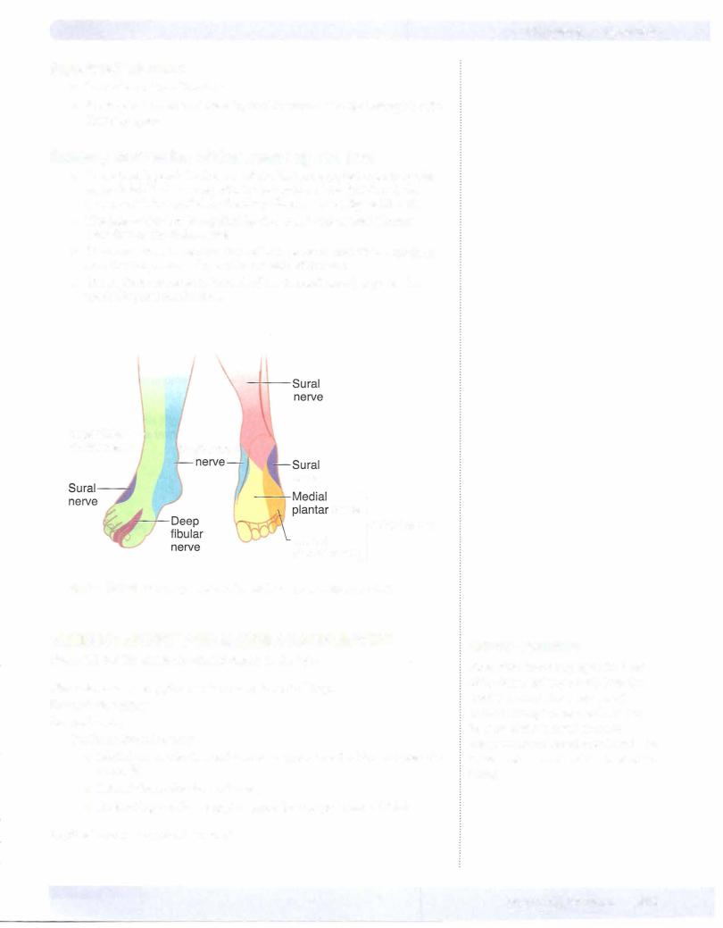
•Sensory loss on the anterior thigh, medial leg, and foot
Obturator Nerve
• Loss ofadduction ofthe thigh as well as sensoryloss on medial thigh
Sciatic Nerve
•Weakened extension ofthe thigh
•Loss of flexion ofthe knee
•Loss ofallfunctions below the knee
•Sensory loss on the posterior thigh, leg (except medial side), and foot
Tibial nerve only
•Weakness in flexion ofthe knee
•Weakness in plantar flexion
•Weakened inversion
•Sensory loss on the leg (except medial) and plantar foot
Commonfibularnerve (neckoffibula)
Produces a combination of deficits oflesions of the deep and superficial fibular nerves
Deepfibularnerve
•Weakened inversion
•Loss ofextension ofthe digits
•Loss of dorsiflexion ("foot drop")
•Sensory loss limited to skin ofthe first web space between the great and second toes