Материал: [2.1] 3D Imaging, Analysis and Applications-Springer-Verlag London (2012)

484 |
P.G. Batchelor et al. |
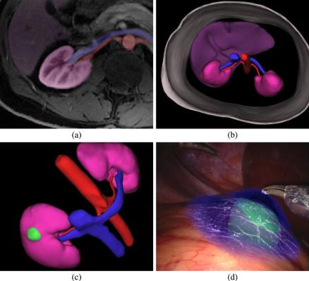
Fig. 11.24 Augmented reality image overlay for partial nephrectomy, showing CT derived model (a), (b), (c) and p − q space rendering (d) of the model superimposed on the surgical view [43, 65]
been used in neurosurgery and orthopaedics where the operation is close to bone, which means that the rigid body registration approximation should hold. To align a preoperative model to the patient, it is necessary for a coordinate system to be established in the operating room. This part of the process can use techniques from computer vision. The most common commercial navigation devices use an optical camera to track markers which are either active, retro-reflective or passive.
An aligned preoperative model can then be displayed using augmented reality [65] to blend the real operative view with the 3D model (see Fig. 11.24).
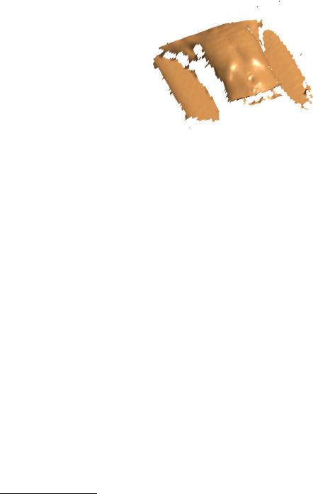
The majority of surface data in medical imaging is extracted from volumetric acquisitions, mostly CT and MRI. However, there is the potential to use surfaces from video sources during therapy or surgery. These surfaces will come from one or more of the techniques described in the other chapters in this book. In radiotherapy, for example, 3D surface reconstruction from projected patterned light is used to track the chest and abdomen position in real-time. This information can be linked with previous volumetric data, such as the CT derived model in Fig. 11.24, to estimate the position of a tumour in real-time, as shown in Fig. 11.25.

486 |
P.G. Batchelor et al. |
In registration, the issue of how to cope with non-rigid soft tissue motion is key to providing accurate alignment. In particular, for image guidance one needs to deform a 3D preoperative model to match the (often 2D) intraoperative scene. The incorporation of physical finite element models into this process helps to keep deformations realistic, but significantly increases the required processing power.
In image segmentation, there is no generic automatic algorithm that has been shown to work reliably. Given the amount of time a clinician must spend doing manual segmentation, more automated approaches are vital if segmentation is to be adopted into routine practice. The segmentation workshop of the MICCAI conference incorporates grand challenges4 for specific clinical applications and algorithms compete in automatic and semi-automatic categories. Although some impressive developments have been made, there is as yet no perfect automated method.
There are some significant unanswered questions in the field of shape modeling. Little attention has been paid to the number of modes that should be retained and what constitutes a sufficient sample. Instead, in most cases, models are built on a limited sample and the number of modes retained uses a simple heuristic such as fitting 95 % of the data. Mei et al. [54, 55] suggest using bootstrapping as a means of assessing the stability of mode directions across replicates. For real and simulated data, the number of modes retained stabilizes at a given sample size and this is taken as an indication of sample sufficiency.
There is also the question of how to establish correspondence. Much work has focused on producing diffeomorphic transformations and the use of the minimum description length to establish optimal correspondences [20]. Other interesting research areas include non-linear models, such as kernel PCA and manifold learning. On a well chosen manifold, the shape model may well be more compact and can be represented by a smaller sample.
Within diffusion tensor imaging, the dominant issues are those surrounding how to generate fibre tracts in the brain or heart. The field of tractography looks at methods to cope with error accumulation and difficult cases, such as fibre tracts that cross each other. There is increasing interest in tractography in neuroscience, since imaging of the connectivity of the brain can potentially help to understand its function.
11.10 Further Reading
As mentioned, the premier conference on medical imaging is MICCAI. The papers in this conference are the best source to find the latest research developments in this field. On a more algorithmic level, IPMI5 provides a smaller but more focussed conference. A superb introduction to medical image processing, with examples in Matlab, is given in [11].
4See www.grand-challenge.org.
5International Conference on Information Processing in Medical Imaging. See www.ipmi-conference.org.

11 3D Medical Imaging |
487 |
Data Acquisition There are many medical physics textbooks and books that specialize in certain modalities. There are too many to give a comprehensive list here, but [76] is a good overview and we can mention a few noteworthy references. The Encyclopedia of Medical Imaging [77] is a good source of information and further references. As a reference on the mathematics of imaging, we would recommend [9]. Readers with an interest in the most mathematical texts should read [57] or [3, 26]. A classic reprint is [39]. For theoretical problems, inspired from tomography but without applications in mind, see [30]. An overview of PET can be found in [1]. For MRI, a signal processing perspective is given in [44] (one of the authors is Paul Lauterbur who was awarded the Nobel prize for inventing MRI).
Surface Extraction The manipulation of surface objects, for example triangulated surfaces, is presented in [25] and gives the basic concepts. For further investigation, again distanced from the medical imaging motivation, see also [12].
Volume Registration For a full review of medical image registration see [35] or the book [31], while the book [74] collects classical papers in information theory.
Segmentation For the latest developments in segmentation visit the ‘grand challenge’ web pages.6 In specific areas, there are some excellent review papers. For deformable models, there is the original paper by McInerney [53]. In statistical shape modeling, an excellent review is given by [34]. Further reviews are available of semi-automatic and fully-automatic segmentation [64], the role of user interaction [59] and ultrasound segmentation [58].
Diffusion Imaging The book [38] is an excellent overview of this application topic. The books [80, 81] contain a range of articles on tensor processing in the spirit of this book.
Image-Guided Surgery An excellent textbook on image guidance is provided by Peters and Cleary [63]. The latest research is also well covered by the conferences IPCAI7 and MICCAI.
Software Medical images come in a number of formats. The standard is DICOM,8 considered clumsy sometimes, but it is the standard that main manufacturers of medical imaging equipment use. Besides manufacturers’ tools, MRIcro, ImageJ, Matlab, and OsiriX are software packages that can read and display them. The most popular open source library is dicom4chee and the C++ libraries vtk and itk also provide lots of tools that can be used as plugins in ImageJ, for example, via Java wrappers. Matlab is commonly used in research environments, in particular in reconstruction
6www.grand-challenge.org.
7International Conference on Information Processing in Computer-Assisted Interventions.
8Digital Imaging and Communications in Medicine.