Материал: 2016_Kaplan_USMLE_Step_1_Lecture_Notes_Pharmacology

The Autonomic Nervous System (ANS) |
1 |
Learning Objectives
Explain information related to anatomy of the ANS
Solve problems concerning blood pressure control mechanisms
Answer questions related to pupillary size and accommodation mechanisms
ANATOMY OF THE ANS
The ANS is the major involuntary portion of the nervous system and is responsible for automatic, unconscious bodily functions, such as control of heart rate and blood pressure and both gastrointestinal and genitourinary functions. The ANS is divided into two major subcategories: the parasympathetic autonomic nervous system (PANS) and the sympathetic autonomic nervous system (SANS).
Location of ANS Ganglia
Both the PANS and SANS have relay stations, or ganglia, between the CNS and the end organ, but the somatic system does not. An important anatomic difference between the SANS and PANS is that the ganglia of the former lie in two paraventral chains adjacent to the vertebral column, whereas most of the ganglia of the PANS system are located in the organs innervated. Figure II-1-1 highlights the major features of the ANS and the somatic systems and also shows the location of the major receptor types. These are:
λ |
NN |
Nicotinic receptors are located on cell bodies in ganglia of |
|
|
both PANS and SANS and in the adrenal medulla. |
λ |
NM |
Nicotinic receptors are located on the skeletal muscle motor |
|
|
end plate innervated by somatic motor nerves. |
λ |
M1–3 |
Muscarinic receptors are located on all organs and tissues |
|
|
innervated by postganglionic nerves of the PANS and on |
|
|
thermoregulatory sweat glands innervated by the SANS. |
39
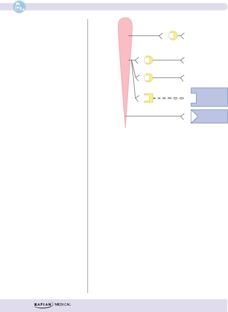
Section II λ Autonomic Pharmacology |
|
|
CNS |
Craniosacral |
PANS |
|
|
Thoraciclumbar |
SANS |
|
|
Postganglionic |
|
|
|
||
|
neuron |
|
|
|
||
Preganglionic |
|
Various organs |
||||
|
|
|
|
|||
neuron |
ACh N |
|
|
|
|
Heart, smooth |
N |
ACh |
M |
|
|||
|
|
muscle, |
||||
|
|
|
|
|
|
|
|
|
|
|
|
|
glands |
|
|
|
|
|
|
|
|
|
|
|
Various organs |
|
ACh N |
|
|
α |
Heart, smooth |
|
N |
NE |
or |
|||
muscle, |
|||||
|
|
|
β |
||
|
|
|
|
glands |
|
|
|
|
|
||
|
|
|
|
|
|
ACh N |
|
|
|
Sweat glands |
|
N |
ACh |
M |
piloerector |
||
|
|
|
|
muscles |
|
|
|
|
|
|
SOMATIC
Neurohumoral |
|
|
Various organs |
transmission |
|
α |
|
ACh NN |
Epi |
or |
Transported |
Adrenal medulla |
|
β |
via blood |
|
|
|
|
Motor neuron |
ACh |
NM |
Skeletal |
|
muscle |
||
Neuromuscular |
|
||
|
|
||
junction |
|
|
|
Figure II-1-1. Anatomy of the Autonomic Nervous System
Bridge to Physiology
For a more detailed discussion, see Section II, Chapter 2, in Physiology.
Neurotransmitters
λAcetylcholine (ACh) is the neurotransmitter at both nicotinic and muscarinic receptors in tissues that are innervated. Note that all direct transmission from the CNS (preganglionic and motor) uses ACh, but postganglionic transmission in the SANS system may use one of the organ-specific transmitters described below.
λNorepinephrine (NE) is the neurotransmitter at most adrenoceptors in organs, as well as in cardiac and smooth muscle.
λDopamine (DA) activates D1 receptors, causing vasodilation in renal and mesenteric vascular beds.
λEpinephrine (E, from adrenal medulla) activates most adrenoceptors and is transported in the blood.
BLOOD PRESSURE CONTROL MECHANISMS
Autonomic Feedback Loop
λBlood pressure is the product of total peripheral resistance (TPR) and cardiac output (CO).
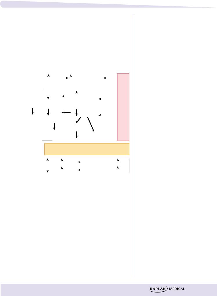
λBoth branches of the ANS are involved in the autonomic (or neural) control of blood pressure via feedback mechanisms.
λChanges in mean blood pressure are detected by baroreceptors, which relay information to the cardiovascular centers in the brainstem controlling PANS and SANS outflow. For example, an increase in mean blood
40
Chapter 1 λ
pressure elicits baroreceptor discharge, resulting in increased PANS activity, leading to bradycardia and decreased SANS activity, which leads, in turn, to decreased heart rate, force of contraction, and vasoconstriction. The resulting decreases in cardiac output and total peripheral resistance
contribute to restoration of mean blood pressure toward its normal level. |
||||||||||||
|
|
|
|
|
|
|
|
|
|
|
r |
|
λ Conversely, decreases in blood pressure elicit ANS neural feedback |
||||||||||||
|
|
|
|
|
|
|
|
|
|
|
l |
|
involving decreased PANS outflow and increased SANS activity— |
||||||||||||
|
|
|
|
|
|
|
|
|
|
|
u |
|
actions leading to increases in cardiac output and total peripheralc |
||||||||||||
resistance. |
|
|
|
|
|
|
|
|
|
|
s |
|
|
|
|
|
|
|
|
|
|
|
a |
||
|
|
|
|
|
|
|
|
|
|
|
v |
|
|
|
|
|
|
|
|
|
|
|
|
o |
|
|
|
|
|
|
|
|
|
|
|
|
i |
|
|
|
|
|
|
|
|
|
|
|
|
d |
|
|
|
|
|
|
|
|
|
|
|
|
r |
|
BP = mean BP |
* |
|
|
|
|
|
baroreceptor |
|
|
|
a |
|
BP |
|
|
|
|
|
C |
||||||
works for either |
|
|
|
|
|
|
discharge |
|
|
|
r |
|
|
|
|
|
|
|
|
|
|
|
e |
||
hyperor hypotension |
|
|
|
|
|
|
|
|
|
|
|
t |
|
|
|
|
|
|
|
|
|
|
|
|
n |
|
|
|
|
|
|
|
|
|
|
|
||
|
|
heart |
|
|
|
|
vagal |
|
|
|
e |
|
|
|
|
|
|
|
|
|
|
||||
|
|
rate |
|
|
|
|
tone |
PANS |
|
C |
||
|
|
|
|
|
|
|
||||||
|
|
|
|
|
|
|
|
|
|
|||
vasoconstriction
B P = T P R ×C O
C O = H R ×S V
Needed for |
|
|
TPR ( |
|
α1) |
|
reflex bradycardia ( |
|
M2) |
|
|
|
|
||||||
|
|||||||||
explanations of |
|
|
TPR ( |
|
β2) |
|
reflex tachycardia ( |
|
β1) |
tracings |
|
|
|
|
|
||||
|
|
|
|
|
|||||
|
|
|
Figure II-1-2. Autonomic Feedback Loop
Hormonal Feedback Loop
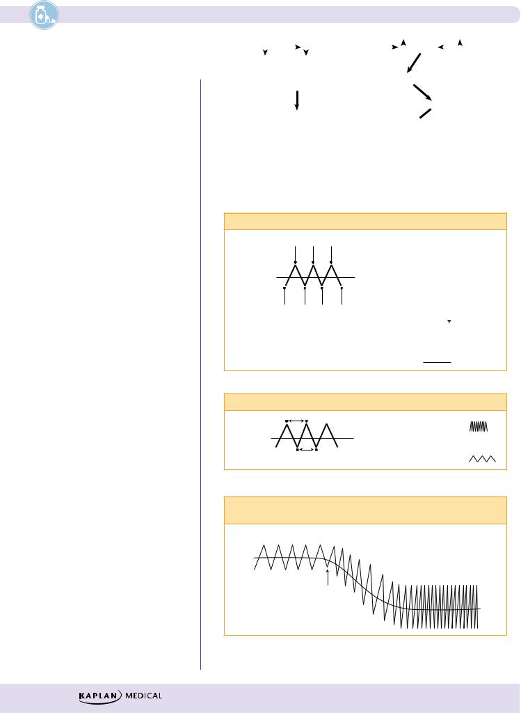
λBlood pressure is also regulated via the hormonal feedback loop shown in Figure II-1-3.
λThe system is affected only by decreases in mean blood pressure (hypotension), which result in decreased renal blood flow.
λDecreased renal pressure causes the release of renin, which promotes formation of the angiotensins.
λAngiotensin II increases aldosterone release from the adrenal cortex, which, via its mineralocorticoid actions to retain sodium and water, increases blood volume.
λIncreased venous return results in an increase in cardiac output.
λAngiotensin II also causes vasoconstriction, resulting in an increase in TPR.
The Autonomic Nervous System (ANS)
Note
Baroreceptor reflexes can be blocked at the ganglionic synapse with NN receptor antagonists. Alternatively, a reflex bradycardia can be blocked with muscarinic antagonists; a reflex tachycardia can be blocked with β1 antagonists.
41
Section II λ Autonomic Pharmacology
Note
Antihypertensive Drugs
Both the ANS (neural) and endocrine feedback loops are invoked when patients are treated with antihypertensive drugs. Such
compensatory mechanisms may result in tachycardia and both salt and water retention.
Works only in |
* |
|
BP |
|
|
Renal blood flow |
|
|
Renin |
|
|
|
Sympathetic |
|
|
|
|
|
|
|
|||||||
|
|
|
|||||||||||
hypotension |
|
|
|
|
|
|
|
|
|
|
|
drive |
|
|
|
|
|
|
|
|
|
|
|
|
|
 Aldosterone
 Angiotensin II
 TPR
TPR
 Blood volume
 CO
CO 
 BP
BP
Figure II-1-3. Hormonal Feedback Loop
Introduction to Blood Pressure/Heart Rate Tracings
Blood Pressure
|
Systolic pressure |
|
BASELINE |
Mean blood |
|
pressure |
||
|
||
|
Diastolic pressure |
•Increases are seen as deflections of the tracing upward
(  )
)
•Decreases are seen as deflections of the tracing downward (  )
)
•Following mean blood pressure changes
is enough
()
A
Heart Rate
|
• Increases are seen as |
( |
) |
BASELINE |
tighter tracing |
|
|
• Decreases are seen as |
|
|
|
|
( |
) |
|
One beat |
a wider tracing |
B
Example of a Drug X Changing Baseline Parameters by Decreasing Mean Blood Pressure and Increasing Heart Rate
BASELINE |
DRUG X EFFECT |
X
C
Figure II-1-4. Blood Pressure/Heart Rate Tracings
42

Chapter 1 λ The Autonomic Nervous System (ANS)
PUPILLARY SIZE AND ACCOMMODATION MECHANISMS
Muscarinic stimulation |
|
Radial muscle (α1) |
Contraction of |
Sphincter muscle (M) |
|
|
sphincter muscle |
Muscarinic stimulation
Normal
Muscarinic
agonist
Miosis
Ciliary |
Contraction of |
||||
muscle (M) |
ciliary muscle |
||||
Suspensory |
|
|
|||
ligament |
|
|
|||
|
Lens |
|
|
|
|
|
|
|
|
||
|
|
|
|
|
|
Normal |
Spasm of |
|
Accommodation |
Adrenergic stimulation Radial muscle (α1)
Sphincter muscle (M)
Adrenergic
stimulation
Normal
Mydriasis |
Contraction of
radial muscle
α1 Agonists
Muscarinic stimulation
1.Miosis
2.Accommodation (near vision)
Muscarinic antagonism
1.Mydriasis
2.Accommodation to far vision, leading to cycloplegia (paralysis of accommodation)
α1-agonists
1.Mydriasis
2.No cycloplegia
Figure II-1-5. Effect of ANS Drugs on the Eye
43