Материал: 2016_Kaplan_USMLE_Step_1_Lecture_Notes_Pharmacology
λDigoxin
−Direct effect: inhibition of cardiac Na+-K+ ATPase
ºResults in ↑ intracellular Na+
º↓ Na+/Ca2+ exchange
º↑ intracellular Ca2+
º↑ Ca2+ release from sarcoplasmic reticulum
º↑ actin-myosin interaction
º↑ contractile force
−Indirect effect: inhibition of neuronal Na+-K+ ATPase
ºResults in ↑ vagal activity
−Pharmacokinetics:
ºLong t1/2: need loading dose (LD)
ºRenal clearance: caution in renal impairment
ºTissue protein binding (large Vd): displacement by other drugs (verapamil, quinidine)
−Uses:
ºCHF
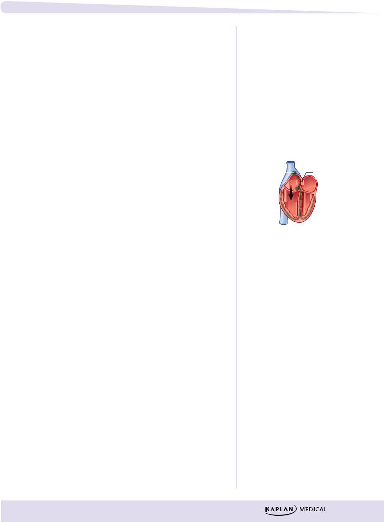
ºSupraventricular tachycardias, except Wolff-Parkinson-White syndrome (see margin note)
−Side effects:
ºEarly signs include anorexia, nausea, ECG changes
ºLater signs include disorientation, visual effects (halos)
ºIn toxic doses, any cardiac arrhythmias
−Management of toxicity
ºUse of Fab antibodies toward digoxin
ºSupportive therapy (electrolytes and antiarrhythmics class IB)
−Drug interactions:
ºDiuretics: ↓ K+, ↓ Mg2+, ↑ Ca2+
ºQuinidine and verapamil
λPhosphodiesterase inhibitors: inamrinone and milrinone
−Use: acute CHF only
−↑ cAMP in heart muscle; results in ↑ inotropy
−↑ cAMP in smooth muscle; results in ↓ TPR
λSympathomimetics: dobutamine and dopamine
−Use: acute CHF only
OTHER DRUGS
−Nesiritide
ºRecombinant form of human B-type natriuretic peptide (rh BNP)
ºBinds to natriuretic peptide receptors, thus ↑ cGMP, resulting in vasodilation
ºUsed in acutely decompensated CHF
Chapter 3 λ Drugs for Heart Failure
Note
Wolff-Parkinson-White Syndrome
SA node |
|
|
AV node |
|
|
||
Conduction |
|
|
|
accessory |
|
(Slow |
|
|
|||
pathways |
|
||
(fast |
|
conduction) |
|
muscle |
|
|
|
fibers) |
|
|
|
λDo:
–block accessory pathway with IA or III
λDon’t:
–slow AV conduction (avoid digoxin, β-blocker, Ca2+-channel blocker, adenosine)
Clinical Correlate
Diastolic dysfunction (CHF with preserved ejection fraction) is best treated with β blockers and diuretics.
99
Section III λ Cardiac and Renal Pharmacology
Chapter Summary
λHeart failure is an inability of the heart to pump with sufficient vigor to maintain an adequate cardiac output. The mechanisms involved are discussed and are illustrated in Figure III-3-1.
λDrugs used to treat heart failure include those that decrease preload (e.g., diuretics, ACEIs, ARBs, and venodilators), those that decrease afterload (e.g., ACEIs, ARBs, and arteriodilators), and those that increase cardiac contractility (e.g., digoxin and beta agonists).
λPrimary treatments for chronic CHF are ACEI, beta blockers, and diuretics.
λDrugs which inhibit cardiac remodeling include ACEI, ARBs, beta blockers, spironolactone, and eplerenone.
λDigoxin enhances cardiac contraction by inducing a series of responses initiated by inhibiting the Na+/K+ ATPase. Figure III-3-2 shows how inhibition of cardiac membrane Na+/K+ ATPase leads to increased contractility.
λDigoxin has potential toxic effects that are in part dependent upon the electrolyte balance.
λBipyridines, sympathomimetics, and nesiritide also have uses in treating acute heart failure.
100
Antiarrhythmic Drugs |
4 |
Learning Objectives
Demonstrate understanding of cardiac action potential
Use knowledge of Na+ channels to explain arrhythmias,
Explain information related to ANS regulation of heart rate
Answer questions about controlling arrhythmias using Na+ channel blockers, beta blockers, K+ channel blockers, Ca2+ channel blockers, and other unclassified drugs
CARDIAC ACTION POTENTIAL
Fast-Response Fibers: Cardiac Muscle, His-Purkinje
System
mV
0
−20
−40
−60
−80
−100
Overshoot (phase 1)
) |
|
Plat |
|
|
|
||||
|
|
|
|
|
e |
|
|
|
|
0 |
( |
|
|
|
a |
u |
|
||
se |
|
|
|
|
|
|
|
||
|
p |
as |
|
|
|
|
|
||
|
|
|
h |
e |
|
|
|
||
pha |
|
|
|
|
2) |
|
|
||
|
Ca |
2+ |
|
|
K |
+ |
|
||
|
|
|
|
||||||
( |
|
|
|
|
|
|
|||
n |
|
|
|
|
|
|
n |
|
|
o |
|
|
|
|
|
|
i |
|
|
ti |
|
|
|
|
|
|
o |
) |
|
|
|
|
|
|
|
t |
|||
za |
|
|
|
|
|
|
|
||
|
|
|
|
|
|
i e |
|||
|
|
Na |
+ |
|
|
|
a |
3 |
|
i |
|
|
|
|
z |
s |
|||
|
|
|
|
|
r |
||||
r |
|
|
|
|
|
|
|
|
|
ola |
|
|
|
|
|
|
a |
|
|
|
|
|
|
|
|
|
|
l a |
|
p |
|
|
|
|
|
|
|
o h |
|
|
|
|
|
|
|
|
p p |
||
e |
|
|
|
|
|
|
|
||
|
|
|
|
|
|
|
e |
( |
|
D |
|
|
|
|
|
|
|
||
|
|
|
|
|
|
|
R |
|
|
|
|
|
|
|
|
|
|
|
|
Fast Na+ current
 K+
K+
Resting potential
(phase 4)
Slow Ca2+ current Delayed rectifier K+ current
Figure III-4-1. Cardiac Action Potentials in Fast-Response Fibers
Phase 0
λNa+ channels open—sodium enters the cell down its concentration gradient (fast INa), causing membrane depolarization.
λRate of depolarization depends on number of Na+ channels open, which in turn depends on resting membrane potential of the cell.
λClass I antiarrhythmic drugs can slow or block phase 0 in fast-response fibers.
101
Section III λ Cardiac and Renal Pharmacology
Phase 1
λNa+ channels are inactivated.
λIn some His-Purkinje cells, transient outward K+ currents and inward Cl– currents contribute to the “notch” and overshoot.
λAntiarrhythmic drugs have no significant effects on these transient currents.
Phase 2
λPlateau phase in which a slow influx of Ca2+ (ICa-L) is “balanced” by a late-appearing outward K+ current (the delayed rectifier current IK).
λAntiarrhythmic drugs have no significant effects on these currents during this phase of the action potential (AP).
Phase 3
λRepolarization phase in which the delayed rectifier K+ current rapidly increases as the Ca2+ current dies out because of time-dependent channel inactivation.
λClass III antiarrhythmic drugs slow this repolarization phase.
λNote that during phases 0 through 3 a slow Na+ current (“window current”) occurs, which can help prolong the duration of the action potential.
Phase 4
λReturn of membrane to resting potential—maintained by activity of the Na+/K+-ATPase.
Responsiveness
λCapacity of a cell to depolarize, associated with the number of Na+ channels in a ready state (see Figure III-4-4).
λThis in turn depends on resting membrane potential: the more negative the resting potential (RP), the faster the response.
Conductance
Rate of spread of an impulse, or conduction velocity—three major determinants:
λRate of phase 0 depolarization—as Vmax decreases, conduction velocity decreases and vice versa.
λThreshold potential—the less negative, the slower the conduction velocity.
λResting potential—the more negative the RP, the faster the conduction.
102
Chapter 4 λ Antiarrhythmic Drugs
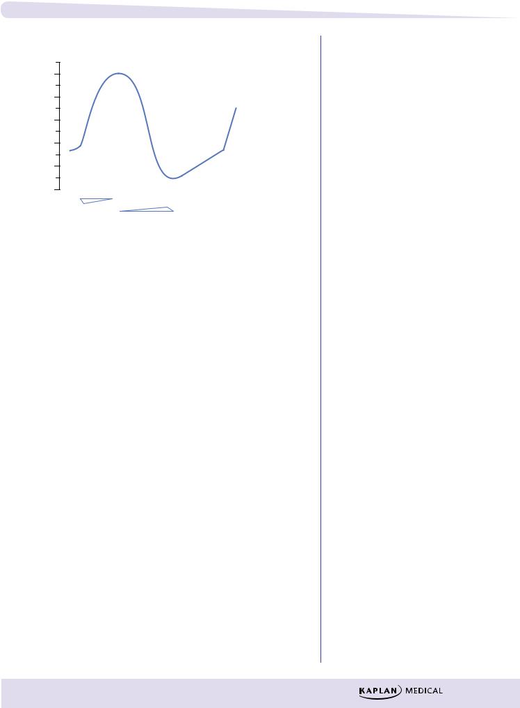
Slow-Response Fibers (SA and AV Nodes, Specialized Cells)
0
−20
−40
−60
−80
−100
↓ K+
↑ Ca2+
Pacemaker
current
↑ Na+, ↑ Ca2+: ↓ K+
Slow Ca+ current
Delayed rectifier K+ current
Figure III-4-2. Cardiac Action Potentials in Slow-Response Fibers
λNo appreciable Na+ current during phase 0 in these cells because the Na channels are either absent or in an inactive form because of the existing voltage.
λDepolarization depends on activation of Ca2+ channels (ICa-L and ICa-T).
λClass IV antiarrhythmic drugs can slow or block phase 0 in slowresponse fibers.
λDuring repolarization, the Ca2+ currents are opposed and overcome by the delayed rectifier K+ current. The relative magnitudes of these opposing currents determine the “shape” of the action potential.
λThe major distinctive feature of slow fibers is their spontaneous depolarization, shown by the rising slope of phase 4 of the AP, referred to
as the pacemaker potential or “pacemaker current.” Although not completely understood, pacemaker potential is a composite of inward Na+ (If) and Ca2+ (ICa-T) currents and outward K+ currents (IK).
λClass II and IV antiarrhythmic drugs can slow phase 4 in pacemaker fibers.
Automaticity
λThe ability to depolarize spontaneously confers automaticity on a tissue.
λThe fastest phase 4 slope will determine the pacemaker of the heart, which is normally the SA node.
Refractoriness
λ The inability to respond to a stimulus—property of all cardiac cells.
103