Материал: 2016_Kaplan_USMLE_Step_1_Lecture_Notes_Pharmacology
Section III λ Cardiac and Renal Pharmacology
Effective Refractory Period (ERP)
λNo stimulus, of any magnitude, can elicit a response.
λLasts into late stage 3 of the AP because Na+ channels are effectively inactivated and not in the “ready” state.
λBlockers of K+ channels prolong the ERP.
Relative Refractory Period (RRP)
λA strong stimulus can elicit a response, but the timing will be out of sync with the rest of the heart and arrhythmias may occur.
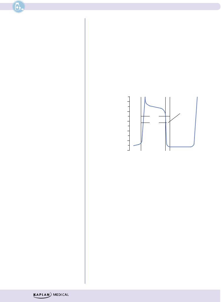
λRatio of ERP to the action potential duration (APD) is a measure of refractoriness, as illustrated in Figure III-4-3. Decreases in ERP favor the formation and propagation of premature impulses.
mV
0
−20
−40
−60
−80
−100
RRP
APD
ERP
Figure III-4-3. Relationship of ERP to APD
104
Chapter 4 λ Antiarrhythmic Drugs
Na+ CHANNELS
Activation
Resting, Ready |
|
Open, Active |
Na+ |
||||||||||
|
Phase 0, Na+ in |
||||||||||||
|
|
|
|
|
|
|
|||||||
|
|
|
|
|
|
|
|
|
|
|
|
|
|
|
|
Closed |
Threshold |
|
|
|
|
Open |
|||||
|
|
|
|
|
|
||||||||
|
|
|
|
|
|
|
|
|
|
||||
|
|
gate opens |
|
|
|
|
|||||||
|
|
|
|
|
|
|
|
|
|
||||
|
|
|
|
|
|
|
|
|
|
|
|
|
|
|
|
|
|
|
|
|
|
|
|
|
|
|
|
|
|
|
Open |
|
|
|
|
|
|
|
Open |
||
|
|
|
|
|
|
|
|
|
|
|
|
|
|
|
|
|
|
|
|
|
|
|
|
|
|
|
|
 Na/K ATPase pump is active.
3 Na out/2 K in, helps repolarization At approx. –50mV ‘M’ gate closes. At approx. –85mV ‘h’ gate opens.
Depolarization of tissue
|
Inactive Refractory |
Repolarization |
Open |
|
|
|
Closed |
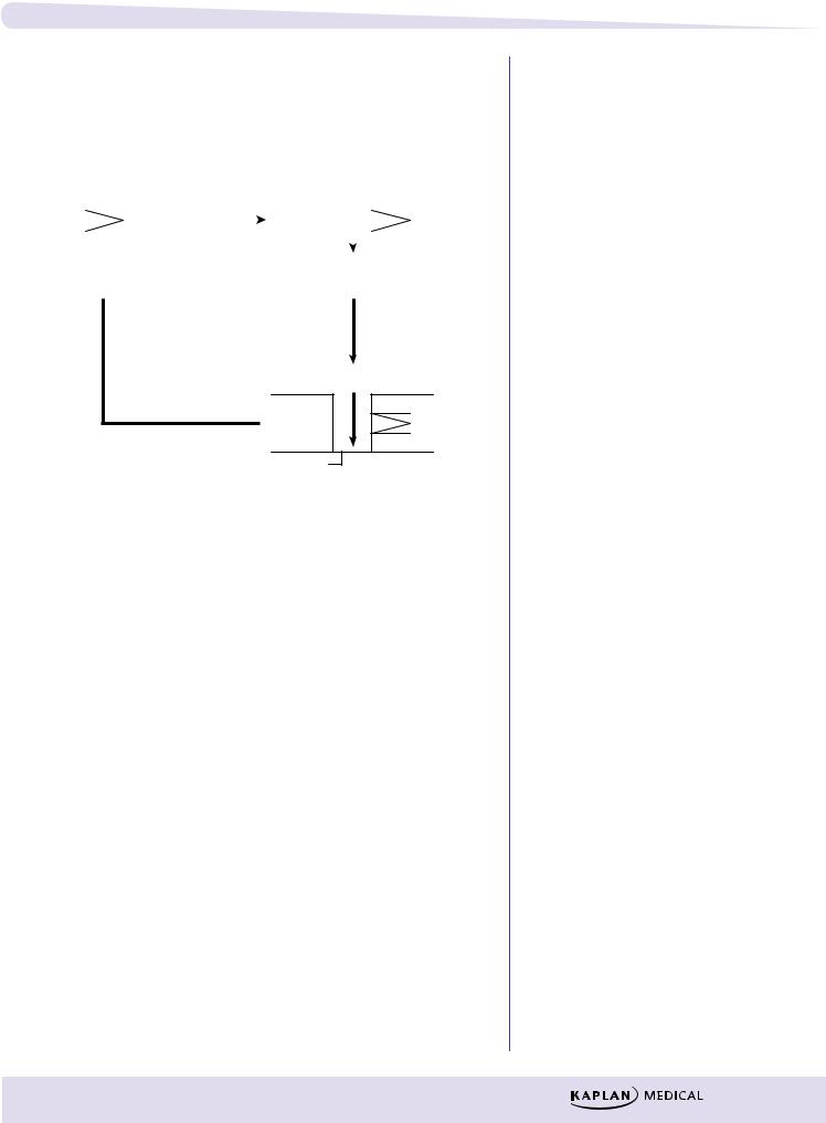
Figure III-4-4. Mechanism of Action of Voltage-Gated Na+ Channels
λThis voltage-gated channel, which is responsible for the fast Na current (INa), exists in three conformations:
–Resting or ready state
–Open or active state
–Inactivated or refractory state
λThe channel has two gates: M (activating) and h (inactivating), both of which are sensitive to voltage changes.
λInactivation of the h gate is slower; therefore, it stays open longer and the Na channel is active.
Recovery
λRate of recovery of the Na channel is dependent on resting potential (RP).
λFastest rate of recovery occurs at normal RP, and recovery slows as membrane voltage increases.
λRate of recovery is slower in ischemic tissue because cells may be partly depolarized at rest. This reduces the number of channels able to participate in the next depolarization, which leads to a decrease in conduction rate in ischemic tissue.
λNa channel blockers also slow the rate of recovery in such tissues.
105

Section III λ Cardiac and Renal Pharmacology
Note
For the exam, you should understand which effect is antiarrhythmic (slows heart) and which is proarrhythmic (speeds up heart).
Note
Quinidine is a weak base, and antacids increase its absorption, thus greatly increasing its toxicity.
ANS REGULATION OF HEART RATE
λNodal tissue, especially that of the SA node, is heavily innervated by both PANS and SANS fibers activating M2 and β1 receptors, respectively.
λPhase 4 slope is increased by an increase in cAMP resulting from β1 receptor activation and slowed by a decrease in cAMP resulting from M2 receptor activation.
λIncrease in cAMP will:
–Increase upstroke velocity in pacemakers by increase of ICa-L
–Shorten AP duration by increase of IK
–Increase HR by increase of If , thus increasing slope of phase 4
λDecrease in cAMP:
–Does the opposite plus produces a K+ current (IK/ACh), which slows the rate of diastolic depolarization and thus decreases HR
–Beta blockers prevent cAMP formation, with primary effects on SA and AV nodal tissues.
CLASS I: Na+ CHANNEL BLOCKERS
Class 1A
λAntiarrhythmic: block fast Na+ channels (↓ INa)
λPreferentially in the open or activated state—“state-dependent” blockade
λ↑ action potential duration (APD) and effective refractory period (ERP)
λAlso blocks K+ channel (prolongs repolarization)
λDrugs:
–Quinidine
ºIn addition to the above, causes muscarinic receptor blockade, which can ↑ HR and AV conduction.
ºMay also cause vasodilation via alpha block with possible reflex tachycardia.
ºOrally effective, wide clinical use in many arrhythmias; in atrial fibrillation, need initial digitalization to slow AV conduction.
ºAdverse effects: cinchonism (GI, tinnitus, ocular dysfunction, CNS excitation), hypotension, prolongation of QRS and ↑ QT interval associated with syncope (torsades).
ºDrug interactions: hyperkalemia enhances effects and vice versa; displaces digoxin from tissue binding sites, enhancing toxicity.
–Procainamide
ºLess muscarinic receptor block
ºMetabolized via N-acetyltransferase (genotypic variation) to N-acetyl procainamide (NAPA), an active metabolite
ºAdverse effects: systemic lupus erythematosus (SLE)–like syndrome (30% incidence) more likely with slow acetylators; hematotoxicity (thrombocytopenia, agranulocytosis); CV effects (torsades)
106

Chapter 4 λ Antiarrhythmic Drugs
Class 1B
λAntiarrhythmic: block fast Na+ channels (↓ INa)
λBlock inactivated channels—preference for tissues partly depolarized (slow conduction in hypoxic and ischemic tissues). This results in an increased threshold for excitation and less excitability of hypoxic heart muscle.
λ↓ APD—due to block of the slow Na+ “window” currents, but this increases diastole and extends the time for recovery.
λDrugs and uses:
−Lidocaine
ºPost-MI
ºOpen-heart surgery
ºDigoxin toxicity
ºSide effects: CNS toxicity (seizures); least cardiotoxic of conventional anti-arrhythmics
ºIV use because of first-pass metabolism
−Mexiletine
ºSame uses as lidocaine
ºOral formulations
Class 1C
λBlock fast Na+ channels (↓ INa), especially His-Purkinje tissue
λNo effect on APD
λNo ANS effects
λDrug:
–Flecainide
ºLimited use because of proarrhythmogenic effects, leading to ↑ in sudden death post-MI and when used prophylactically in VT
CLASS II: BETA BLOCKERS
λPrevent β-receptor activation, which would normally ↑ cAMP
λ↓ SA and AV nodal activity
λ↓ Slope of phase 4 (diastolic currents) of AP in pacemakers
λDrugs:
–Propranolol (nonselective) and the cardioselective drugs: acebutolol and esmolol
–Uses:
ºProphylaxis post-MI and in supraventricular tachyarrhythmias (SVTs)
ºEsmolol (IV) is used in acute SVTs
107

Section III λ Cardiac and Renal Pharmacology
Clinical Correlate
Long QT Syndrome
A familial condition associated with increased risk of ventricular arrhythmias may result from mutation in the gene encoding cardiac potassium channels. Class IA and class III antiarrhythmic drugs may increase the risk of torsades in such patients.
Treatment of Torsade
λCorrect hypokalemia.
λCorrect hypomagnesemia.
λDiscontinue drugs that prolong the QT interval.
Clinical Correlate
Atrial fibrillation is the most common arrhythmia in the United States. The primary goals for treatment are:
1.ventricular rate control with beta blockers, CCBs, or digoxin; and
2.anticoagulation.
Clinical Correlate
Potassium
Both hyperkalemia and hypokalemia are arrhythmogenic.
CLASS III: K+ CHANNEL BLOCKERS
λ↓ IK (delayed rectifier current) slowing phase 3 (repolarization) of AP
λ↑ APD and ERP, especially in Purkinje and ventricular fibers
λDrugs:
−Amiodarone
ºMimics classes I, II, III, and IV
ºIncrease APD and ERP in all cardiac tissues
ºUses: any arrhythmias
ºt1/2 >80 days
ºBinds extensively to tissues (large Vd and multiple effects)
ºSide effects:
Pulmonary fibrosis
Blue pigmentation of the skin (“smurf skin”)
Phototoxicity
Corneal deposits
Hepatic necrosis
Thyroid dysfunction
−Sotalol:
º↓ IK, slowing phase III
ºNon-selective beta blocker: β1 blockade, leading to ↓ HR, ↓ AV conduction
ºUse: life-threatening ventricular arrhythmia
ºSide effects: torsades
CLASS IV: Ca2+ CHANNEL BLOCKERS
λBlock slow cardiac Ca2+ channels
λ↓ phase 0, ↓ phase 4
λ↓ SA, ↓ AV nodal activity
λDrugs:
−Verapamil and diltiazem
ºPrototype Ca2+-channel blockers (see Antihypertensive Drugs and Antianginal Drugs chapters in this section)
ºUses: supraventricular tachycardias
ºSide effects: constipation (verapamil), dizziness, flushing, hypotension, AV block
ºDrug interaction:
Additive AV block with β-blockers, digoxin
Verapamil displaces digoxin from tissue-binding sites
UNCLASSIFIED
λAdenosine
−Activates adenosine receptors: causes Gi-coupled decrease in cAMP
−↓ SA and AV nodal activity
108