Материал: 2019 ESC - supraventricular tachycardia
6 |
|
|
|
|
|
|
ESC Guidelines |
|
|
|
|||||
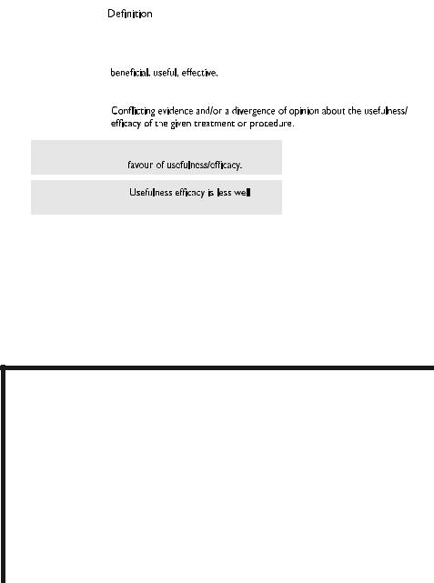
Table 1 Classes of recommendations |
|
|
|||||
|
|
|
|
|
|
Wording to use |
|
recommendationsof |
|
|
|
|
|
|
|
|
|
|
|
|
|
|
|
|
|
|
|
|
|
|
|
|
|
Class I |
Evidence and/or general agreement |
Is recommended or is indicated |
|
||
|
|
|
|
||||
|
|
|
|
that a given treatment or procedure is |
|
|
|
|
|
|
|
|
|
|
|
|
|
|
|
|
|
|
|
|
|
|
Class II |
|
|
|
|
Classes |
|
|
|
|
|
|
|
|
|
|
|
|
|
|
|
|
|
Class IIa |
|
Weight of evidence/opinion is in |
Should be considered |
|
|
|
|
|
|
|
|||
|
|
|
|
|
|
|
|
|
|
|
|
|
|
|
|
|
|
|
Class IIb |
|
established by evidence/opinion. |
May be considered |
|
|
|
|
|
|
|
|
|
|
|
|
|
|
|
|
|
|
|
|
Class III |
Evidence or general agreement that the |
Is not recommended |
2019 |
|
|
|
|
|
given treatment or procedure is not |
|
||
|
|
|
|
useful/effective, and in some cases |
|
© ESC |
|
|
|
|
|
may be harmful. |
|
||
|
|
|
|
|
|
|
|
Table 2 Classes of recommendations
Level of |
|
Data derived from multiple randomized clinical trials |
|
evidence A |
|
or meta-analyses. |
|
|
|
|
|
|
|
|
|
Level of |
|
Data derived from a single randomized clinical trial |
|
evidence B |
|
or large non-randomized studies. |
|
|
|
|
|
|
|
|
|
Level of |
|
Consensus of opinion of the experts and/or small studies, |
2019 |
|
ESC© |
||
evidence C |
|
retrospective studies, registries. |
|
|
|
||
|
|
|
|
slides, booklets with essential messages, summary cards for nonspecialists and an electronic version for digital applications (smartphones, etc.). These versions are abridged and thus, for more detailed information, the user should always access to the full text version of the Guidelines, which is freely available via the ESC website and hosted on the EHJ website. The National Societies of the ESC are encouraged to endorse, translate and implement all ESC Guidelines. Implementation programmes are needed because it has been shown that the outcome of disease may be
.
.
.
.
.
.
.
.
.
.
.
.
.
.
.
.
.
.
.
.
.
.
.
.
.
.
.
favourably influenced by the thorough application of clinical recommendations.
Health professionals are encouraged to take the ESC Guidelines fully into account when exercising their clinical judgment, as well as in the determination and the implementation of preventive, diagnostic or therapeutic medical strategies. However, the ESC Guidelines do not override in any way whatsoever the individual responsibility of health professionals to make appropriate and accurate decisions in consideration of each patient’s health condition and in consultation
2019 September 13 on guest by 1093/eurheartj/ehz467/5556821.abstract/doi/10-article-com/eurheartj/advance.oup.https://academic from Downloaded

ESC Guidelines |
7 |
|
|
with that patient or the patient’s caregiver where appropriate and/or necessary. It is also the health professional’s responsibility to verify the rules and regulations applicable in each country to drugs and devices at the time of prescription.
2 Introduction
Supraventricular arrhythmias are common and patients are often symptomatic, requiring management with drugs and electrophysiological procedures. The ESC published management Guidelines for supraventricular tachycardias (SVTs) in 20031; corresponding US guidelines have also been published, the most recent being in 2015.2
There is a need to provide expert recommendations for professionals participating in the care of patients presenting with SVT. In addition, several associated conditions where SVTs may coexist need to be explained in more detail. To address this topic, a Task Force was convened by the ESC, with the remit to comprehensively review the published evidence and to publish Guidelines on the management of patients with SVT, with up-to-date consensus recommendations for clinical practice. This document summarizes current developments in the field, with a focus on advances since the last ESC Guidelines were published, and provides general recommendations for the management of adults with SVT based on the principles of evidence-based medicine.
2.1 Evidence review
Members of the Task Force were asked to perform a detailed literature review, weigh the strength of evidence for or against a particular treatment or procedure, and include estimates of expected health outcomes where data exist. Patient-specific modifiers, comorbidities, and issues of patient preference that might influence the choice of particular tests or therapies were considered, as were frequency of follow-up and cost-effectiveness. In controversial areas, or with regard to issues without evidence other than usual clinical practice, consensus was achieved by agreement of the expert panel after thorough deliberation. The document was peer-reviewed by official external reviewers.
The strengths of the recommendations and levels of evidence of particular management options were weighed and graded according to predefined scales, as outlined above in Tables 1 and 2, respectively.
Overall, these Guidelines include evidence and expert opinions from several countries. The pharmacological and nonpharmacological antiarrhythmic approaches discussed may therefore include drugs that do not have the approval of governmental regulatory agencies in all countries.
2.2 Relationships with industry and other conflicts of interest
It is ESC policy to sponsor position papers and Guidelines without commercial support, and all members volunteered their time. Thus, all members of the writing group, as well as the reviewers, have disclosed any potential conflicts of interest in detail. The details of these declarations will be posted on the ESC website upon publication of these Guidelines.
.
.
.
.
.
.
.
.
.
.
.
.
.
.
.
.
.
.
.
.
.
.
.
.
2.3 What is new in the 2019 Guidelines?
2.3.1 Change in recommendations from 2003 to 2019
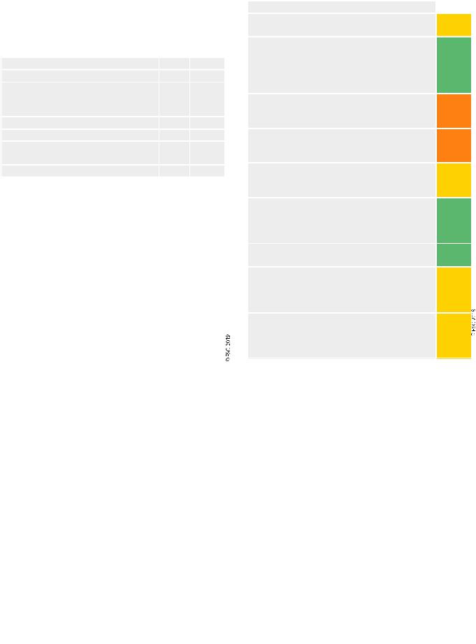
Owing to the fact that the previous Guidelines on SVT were published 16 years ago, many drugs that were recommended at that time are not considered in the 2019 Guidelines. In addition, techniques and indications of ablation techniques have changed. These recommendation changes are indicated for each section in Table 3 below.
Table 3 Changes in SVT Guidelines recommendations since 2003
|
2003 |
2019 |
Acute management of narrow QRS |
|
|
tachycardias |
|
|
Verapamil and diltiazem |
I |
IIa |
Beta-blockers |
IIb |
IIa |
Amiodarone and digoxin are not mentioned in the |
|
|
2019 Guidelines |
|
|
Acute management of wide QRS |
|
|
tachycardias |
|
|
Procainamide |
I |
IIa |
Adenosine |
IIb |
IIa |
Amiodarone |
I |
IIb |
Sotalol and lidocaine are not mentioned in the |
|
|
2019 Guidelines |
|
|
|
|
|
Therapy of inappropriate sinus |
|
|
tachycardia |
|
|
Beta-blockers |
I |
IIa |
Verapamil/diltiazem and catheter ablation are not |
|
|
mentioned in the 2019 Guidelines |
|
|
Therapy of postural orthostatic tachy- |
|
|
cardia syndrome |
|
|
Salt and fluid intake |
IIa |
IIb |
Head-up tilt sleep, compression stockings, selective |
|
|
beta-blockers, fludrocortisone, clonidine, methyl- |
|
|
phenidate, fluoxetine, erythropoietin, ergotamine/ |
|
|
octreotide, and phenobarbitone are not mentioned |
|
|
in the 2019 guidelines. |
|
|
|
|
|
Therapy of focal AT |
|
|
Acute |
|
|
Flecainide/propafenone |
IIa |
IIb |
Beta-blockers |
I |
IIa |
Amiodarone |
IIa |
IIb |
Procainamide, sotalol, and digoxin are not men- |
|
|
tioned in the 2019 Guidelines |
|
|
Chronic |
|
|
Beta-blockers |
I |
IIa |
|
|
|
Verapamil and diltiazem |
I |
IIa |
Amiodarone, sotalol, and disopyramide are not |
|
|
mentioned in the 2019 Guidelines |
|
|
Therapy of atrial flutter |
|
|
Acute |
|
|
Atrial or transoesophageal pacing |
I |
IIb |
Ibutilide |
IIa |
I |
Flecainide/propafenone |
IIb |
III |
|
|
Continued |
2019 September 13 on guest by 1093/eurheartj/ehz467/5556821.abstract/doi/10-article-com/eurheartj/advance.oup.https://academic from Downloaded
8 |
ESC Guidelines |
|
|
Table 3 Continued
|
|
2003 |
2019 |
|
Verapamil and diltiazem |
I |
IIa |
|
|
|
|
|
Beta-blockers |
I |
IIa |
|
Digitalis is not mentioned in the 2019 Guidelines |
|
|
|
Chronic |
|
|
|
Dofetilide, sotalol, flecainide, propafenone, procai- |
|
|
|
namide, quinidine, and disopyramide are not men- |
|
|
|
tioned in the 2019 Guidelines |
|
|
|
Therapy of AVNRT |
|
|
|
Acute |
|
|
|
Amiodarone, sotalol, flecainide, and propafenone |
|
|
|
are not mentioned in the 2019 Guidelines |
|
|
|
Chronic |
|
|
|
|
|
|
|
Verapamil and diltiazem |
I |
IIa |
|
Beta-blockers |
I |
IIa |
|
Amiodarone, sotalol, flecainide, propafenone, and |
|
|
|
the ‘pill-in-the pocket’ approach are not men- |
|
|
|
tioned in the 2019 Guidelines |
|
|
|
Therapy of AVRT |
|
|
|
Flecainide/propafenone |
IIa |
IIb |
|
Beta-blockers |
IIb |
IIa |
|
Amiodarone, sotalol, and the ‘pill-in-the pocket’ |
|
|
|
approach are not mentioned in the 2019 |
|
|
|
Guidelines |
|
|
|
SVT in pregnancy |
|
|
|
Verapamil |
IIb |
IIa |
|
Catheter ablation |
IIb |
IIa* |
|
Sotalol, propranolol, quinidine, and procainamide |
|
|
|
are not mentioned in the 2019 Guidelines. |
|
|
|
|
|
|
*: when fluoroless ablation is available. AT = atrial tachycardia; AVNRT = atrioventricular nodal re-entrant tachycardia; AVRT = atrioventricular re-entrant tachycardia.
i.v. amiodarone is not recommended for pre-excited AF. |
III |
|
|
Performance of an EPS to risk-stratify individuals with
IIa
asymptomatic pre-excitation should be considered.
Catheter ablation is recommended in asymptomatic patients in whom electrophysiology testing with the
use of isoprenaline identifies high-risk properties, such I as SPERRI <250 ms, AP ERP <250 ms, multiple APs,
and an inducible AP-mediated tachycardia.
Non-invasive evaluation of the conducting properties
of the AP in individuals with asymptomatic pre-excita- IIb tion may be considered.
Catheter ablation may be considered in a patient with
asymptomatic pre-excitation and low-risk AP at invaIIb sive or non-invasive risk stratification.
Catheter ablations should be considered in patients
with asymptomatic pre-excitation and LV dysfunction IIa due to electrical dyssynchrony.
AV nodal ablation with subsequent pacing (‘ablate and
pace’), either biventricular or His-bundle pacing, is
I
recommended if a tachycardia responsible for TCM cannot be ablated or controlled by drugs.
During the first trimester of pregnancy, it is recommended
I
that all antiarrhythmic drugs are avoided, if possible.
In pregnant women, beta-1 selective blockers (except
atenolol) or verapamil, in order of preference, should
IIa
be considered for prevention of SVT in patients without WPW syndrome.
In pregnant women, flecainide or propafenone should
be considered for prevention of SVT in patients with
IIa
WPW syndrome and without ischaemic or structural heart disease.
AF = atrial fibrillation; AP = accessory pathway; AT = atrial tachycardia; AV = atrioventricular; EPS = electrophysiology study; ERP = effective refractory period; i.v. = intravenous; LV = left ventricular; POTS: postural orthostatic tachycardia syndrome; SPERRI = shortest pre-excited RR interval during atrial fibrillation; SVT = supraventricular tachycardia; TCM = tachycardiomyopathy; WPW = Wolff-Parkinson-White.
2.3.2 New recommendations in 2019
Table 4 New recommendations in 2019
Ivabradine alone or in combination with a beta- |
|
|
blocker should be considered in symptomatic patients |
IIa |
|
with inappropriate sinus tachycardia. |
|
|
Ibutilide (i.v.) ibutilide may be considered for acute |
IIb |
|
therapy of focal atrial tachycardia. |
||
|
||
Ivabradine for postural orthostatic tachycardia syn- |
|
|
drome, and ivabradine with a beta-blocker for chronic |
IIb |
|
therapy of focal atrial tachycardia, may be considered. |
|
|
Patients with atrial flutter without AF should be con- |
|
|
sidered for anticoagulation, but the threshold for ini- |
IIa |
|
tiation is not established. |
|
|
Ibutilide (i.v.), or i.v. or oral (in-hospital) dofetilide are |
I |
|
recommended for conversion of atrial flutter. |
||
|
||
High-rate atrial pacing is recommended for termina- |
|
|
tion of atrial flutter in the presence of an implanted |
I |
|
pacemaker or defibrillator. |
|
|
|
Continued |
.
.
.
.
.
.
.
.
.
.
.
.
.
.
.
.
.
.
.
.
.
.
.
.
.
.
.
.
.
.
.
.
.
.
.
.
.
.
.
.
.
.
.
.
.
.
.
.
.
.
.
.
.
.
.
.
.
.
2.3.3 New revised concepts
•Drug therapy for inappropriate sinus tachycardia and focal atrial tachycardia.
•Therapeutic options for acute conversion and anticoagulation of atrial flutter.
•Therapy of atrioventricular nodal re-entrant tachycardia.
•Therapy of antidromic atrioventricular re-entrant tachycardia and pre-excited atrial fibrillation.
•Management of patients with asymptomatic pre-excitation.
•Diagnosis and therapy of tachycardiomyopathy.
3 Definitions and classification
The term ‘SVT’ literally indicates tachycardia [atrial rates >100 beats per minute (b.p.m.) at rest], the mechanism of which involves tissue from the His bundle or above.2,3 Traditionally, SVT has been used to describe all kinds of tachycardias apart from ventricular tachycardias (VTs) and AF. It has therefore included tachycardias such as atrioventricular (AV) re-entry due to accessory connections, which is not, in
2019 September 13 on guest by 1093/eurheartj/ehz467/5556821.abstract/doi/10-article-com/eurheartj/advance.oup.https://academic from Downloaded
ESC Guidelines |
9 |
|
|
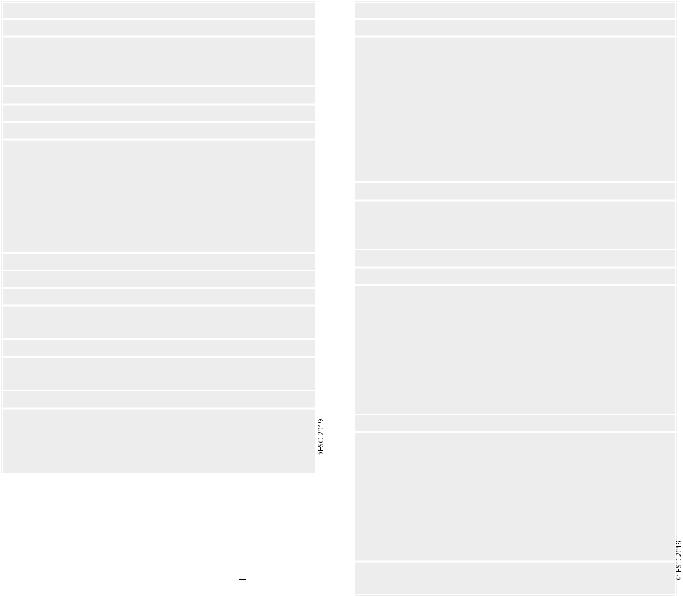
Table 5 Conventional classification of supraventricular |
Table 6 Differential diagnosis of narrow and wide QRS |
tachycardias |
tachycardias |
Atrial tachycardias
Sinus tachycardia
•Physiological sinus tachycardia
•Inappropriate sinus tachycardia
•Sinus nodal re-entrant tachycardia
Focal AT
Multifocal AT
MRAT
•Cavotricuspid isthmus-dependent MRAT
Typical atrial flutter, counter-clockwise (common) or clockwise (reverse)
Other cavotricuspid isthmus-dependent MRAT
•Non-cavotricuspid isthmus-dependent MRAT
RA MRAT
LA MRAT
AF
AV junctional tachycardias
Atrioventricular nodal re-entrant tachycardia (AVNRT)
•Typical
•Atypical
Non-re-entrant junctional tachycardia
•JET (junctional ectopic or focal junctional tachycardia)
•Other non-re-entrant variants
Atrioventricular re-entrant tachycardia (AVRT)
•Orthodromic (including PJRT)
•Antidromic (with retrograde conduction through the AVN or, rarely, over another pathway)
AF = atrial fibrillation; AT = atrial tachycardia; AV = atrioventricular; AVN = atrioventricular node; JET = junctional ectopic tachycardia; RA = right atrial; LA = left atrial; MRAT = macro-re-entrant atrial tachycardia; PJRT = permanent junctional reciprocating tachycardia; RA = right atrial.
essence, a supraventricular rhythm (Table 5). The term ‘narrow QRS tachycardia’ indicates those with a QRS duration <120 ms. A wide
QRS tachycardia refers to one with a QRS duration >120 ms (Table 6). In clinical practice, SVT may present as narrow or wide QRS tachycardias, most of which, although not invariably, manifest as regular rhythms. These Guidelines do not cover AF, which is the subject of separate clinical Guidelines4 and various consensus documents.5 7
4 Electrophysiological mechanisms of supraventricular tachycardia
Arrhythmia can originate from abnormal impulse initiation in an individual myocyte or, more realistically, in a close cluster of myocytes. This can occur in non-pacemaker cells through a mechanism similar to the physiological automaticity of pacemaker cells [sinus node and AV node (AVN)], and is thus named ‘abnormal’ or ‘enhanced automaticity’. An alternative form of abnormal impulse initiation involves oscillations of
Narrow QRS ( 120 ms) tachycardias
Regular
•Physiological sinus tachycardia
•Inappropriate sinus tachycardia
•Sinus nodal re-entrant tachycardia
•Focal AT
•Atrial flutter with fixed AV conduction
•AVNRT
•JET (or other non-re-entrant variants)
•Orthodromic AVRT
•Idiopathic VT (especially high septal VT)
Irregular
•AF
•Focal AT or atrial flutter with varying AV block
•Multifocal AT
Wide QRS (>120 ms) tachycardias
Regular
•VT/flutter
•Ventricular paced rhythm
•Antidromic AVRT
•SVTs with aberration/BBB (pre-existing or rate-dependent during tachycardia)
•Atrial or junctional tachycardia with pre-excitation/bystander AP
•SVT with QRS widening due to electrolyte disturbance or antiarrhythmic drugs
Irregular
•AF or atrial flutter or focal AT with varying block conducted with aberration
•Antidromic AV re-entrant tachycardia due to a nodo-ventricular/fascic- ular AP with variable VA conduction
•Pre-excited AF
•Polymorphic VT
•Torsade de pointes
•Ventricular fibrillation
Occasionally, AF with very fast ventricular response may apparently resemble a regular narrow QRS tachycardia.
AF = atrial fibrillation; AP = accessory pathway; AT = atrial tachycardia; AV = atrioventricular; AVNRT = atrioventricular nodal re-entrant tachycardia; AVRT = atrioventricular re-entrant tachycardia; BBB = bundle branch block; JET = junctional ectopic tachycardia; SVT = supraventricular tachycardia; VA = ventriculoatrial; VT = ventricular tachycardia.
. |
membrane potential, named early or delayed ‘after-depolarizations’. In |
|||||
. |
||||||
. |
|
|
|
|
|
|
. |
such cases, the resulting arrhythmias take the name of ‘triggered activ- |
|||||
. |
||||||
. |
8 |
|
|
|
|
|
. |
|
|
|
|
|
|
. |
ity’. Arrhythmias resulting from enhanced automaticity and triggered |
|||||
. |
|
|
|
|
|
|
. |
activity |
are defined as ‘non-re-entrant’. Arrhythmias can |
also arise |
|||
. |
||||||
. |
|
|
|
|
|
|
. |
when |
myocardial |
regions |
activated |
later in propagation |
re-excite |
. |
||||||
. |
|
|
|
|
|
|
. |
regions that have |
already |
recovered |
excitability. This results from |
||
. |
||||||
. |
|
|
|
|
|
|
. |
abnormal propagation of the excitation wavefront and/or of tissue |
|||||
. |
||||||
. |
||||||
. |
refractoriness. This mechanism, named ‘re-entry’, is based on the syn- |
|||||
. |
||||||
. |
||||||
. |
|
|
|
|
|
|
. |
cytial nature of myocardial tissue and is thus radically different from |
|||||
. |
||||||
. |
|
|
8 |
|
|
|
. |
focal impulse initiation. A detailed discussion and schematic represen- |
|||||
. |
||||||
. |
|
|
|
|
|
|
. |
tation of common SVT circuits is provided in the Supplementary Data. |
|||||
. |
||||||
2019 September 13 on guest by 1093/eurheartj/ehz467/5556821.abstract/doi/10-article-com/eurheartj/advance.oup.https://academic from Downloaded

10 |
ESC Guidelines |
5 Cardiac anatomy for the electrophysiologist
Knowledge of anatomical structures inside and outside the atrial chambers is of clinical importance, especially when interventional procedures are being considered. A detailed discussion is provided in the Supplementary Data.
6 Epidemiology of supraventricular tachycardia
Epidemiological studies on the SVT population are limited. In the general population, the SVT prevalence is 2.25/1000 persons and the incidence is 35/100 000 person-years. Women have a risk of developing SVT that is two times greater than that of men, and persons aged >65 years or have more than five times the risk of developing SVT than younger individuals. Patients with lone paroxysmal SVT vs. those with cardiovascular disease are younger, have a faster SVT rate, have an earlier onset of symptoms, and are more likely to have their condition first documented in the emergency department.9 In a paediatric cohort study of 1 967 911 live births between 2000 08, 2021 patients (51.6% male, overall incidence 1.03/1000) had SVT [Wolff-Parkinson-White (WPW) syndrome accounted for 16.2%]. By the age of 15 years, the annual risk of sudden death was 0.01% per patient-year.10
In specialized centres, AVNRT is the most frequently treated substrate after AF, followed by atrial flutter and AVRT, in patients referred for catheter ablation.11 13 Women are more likely to be affected by AVNRT than men (ratio 70:30),14 16 while the converse is true for AVRT (ratio 45:55).14 A relationship with the monthly cycle has been suggested,17 and episodes are more frequent during pregnancy in women with pre-existing SVT.18
In the general population, the prevalence of a WPW pattern on surface electrocardiogram (ECG) ranges from 0.15 0.25%,19 increasing to 0.55% among first-degree relatives of affected patients.20 However, not all patients develop SVT and intermittent pre-excitation is not rare. Compared with the remaining population, the pre-excitation population is generally younger, predominantly male, and has less comorbidity.21,22 The proportion of patients with AVRT decreases with age, whereas the proportion of those with AVNRT and AT increases with age.14
The epidemiology of atrial flutter is not known with certainty, as atrial flutter and AF can coexist. The prevalence of AF before flutter ablation ranges from 24 62%, and after ablation could be 30 70%. The overall incidence of atrial flutter is 88/100 000 person-years in the US population annually. Adjusted for age, the incidence of atrial flutter in men (125/100 000) is >2.5 times that of women (59/ 100 000) and increases exponentially with age. Patients with atrial flutter are more likely to have been smokers, have a longer PR interval, history of myocardial infarction (MI), and history of heart failure (HF).23
Catheter ablation is now used extensively for most varieties of SVT, and patient-reported outcome measures have shown that patients experience significant improvements in their quality of life following ablation.24 27 Patient-reported outcome measures using
. |
|
|
|
|
|
|
|
|
|
|
|
|
|
|
. |
|
|
|
|
|
|
|
|
|
|
|
|
|
|
. various questionnaires are useful in the audit of ablation techniques. |
||||||||||||||
. |
|
|
|
|
|
|
|
|
|
|
|
|
|
|
. |
Women are more often prescribed antiarrhythmic drugs before abla- |
|||||||||||||
. |
||||||||||||||
. |
|
|
|
28 |
|
|
|
|
|
|
|
|
|
|
. |
|
|
|
and recurrence rates following AVNRT |
||||||||||
. |
tion for SVT than men, |
|||||||||||||
. |
|
|
|
|
|
|
29 |
|
|
|
|
|
|
|
. |
|
|
|
|
|
|
However, overall, no signifi- |
|||||||
. |
ablation are higher in young women. |
|
||||||||||||
. |
|
|
|
|
|
|
|
|
|
|
|
|
|
|
. |
cant differences in health-related quality of life or access to healthcare |
|||||||||||||
. |
||||||||||||||
. |
|
|
|
|
|
|
|
|
|
|
|
|
28 |
|
. |
|
|
|
|
|
|
|
|
|
|
|
|
||
. |
resources between men and women have been reported. |
|
||||||||||||
. |
|
|
|
|
|
|
|
|
|
|
|
|
|
|
. |
|
|
|
|
|
|
|
|
|
|
|
|
|
|
. |
|
|
|
|
|
|
|
|
|
|
|
|
|
|
. |
|
|
|
|
|
|
|
|
|
|
|
|
|
|
. |
|
|
|
|
|
|
|
|
|
|
|
|
|
|
. |
|
|
|
|
|
|
|
|
|
|
|
|
|
|
. |
|
|
|
|
|
|
|
|
|
|
|
|
|
|
. |
7 Clinical presentation |
|
|
|
|
|||||||||
. |
|
|
|
|
||||||||||
. |
|
|
|
|
|
|
|
|
|
|
|
|
|
|
. |
|
|
|
|
|
|
|
|
|
|
|
|
|
|
. |
|
|
|
|
|
|
|
|
|
|
|
|
|
|
. |
The impact of SVT on an individual is based on a range of factors and |
|||||||||||||
. |
||||||||||||||
. |
|
|
|
|
|
|
|
|
|
|
|
|
|
|
. |
may result in palpitations, fatigue, light-headedness, chest discomfort, |
|||||||||||||
. |
||||||||||||||
. |
|
|
|
|
|
|
|
30 |
|
|
|
|
|
|
. |
dyspnoea, and altered |
|
|
|
|
Rapid rhythms |
are more |
|||||||
. |
consciousness. |
|
||||||||||||
. |
|
|
|
|
|
|
|
|
|
|
|
|
|
|
. |
likely to present |
with |
a |
clear history |
and |
acutely than |
less-rapid |
|||||||
. |
||||||||||||||
. |
|
|
|
|
|
|
|
|
|
|
|
31 |
|
|
. |
|
|
|
|
|
|
|
|
|
|
|
|
||
. |
rhythms, but SVT usually produces symptoms. |
The duration of |
||||||||||||
. |
|
|
|
|
|
|
|
|
|
|
|
|
|
|
. |
symptoms and the patient’s age at onset are important. An individual |
|||||||||||||
. |
||||||||||||||
. |
|
|
|
|
|
|
|
|
|
|
|
|
|
|
. |
with an onset in the teenage years or younger is less likely to have AT |
|||||||||||||
. |
||||||||||||||
. |
|
|
|
|
|
|
|
|
|
|
|
|
|
|
. |
or AF continuing into |
adulthood. Such a |
long |
history |
will point |
|||||||||
. |
||||||||||||||
. |
|
|
|
|
|
|
|
|
|
|
|
|
|
|
. |
|
|
|
|
32 |
Dyspnoea, or other clinical signs |
||||||||
. |
towards a re-entrant mechanism. |
|||||||||||||
. |
|
|
|
|
|
|
|
|
|
|
|
|
|
|
. |
and symptoms of HF, can occur when the patient has developed |
|||||||||||||
. |
||||||||||||||
. |
||||||||||||||
. |
|
|
|
|
|
|
|
|
|
|
|
|
|
30 |
. |
TCM. Light-headedness in association with SVT is not infrequent. |
|||||||||||||
. |
||||||||||||||
. |
|
|
|
|
|
|
|
|
|
33,34 |
|
|
|
|
. |
|
|
|
|
|
|
|
|
|
and tend to be asso- |
||||
. |
Presyncope and syncope are less common, |
|
||||||||||||
. |
|
|
|
|
|
|
|
|
|
|
|
32 |
|
|
. |
ciated with |
presentation |
in older |
|
individuals. |
|
In older patients, |
|||||||
. |
|
|
||||||||||||
. |
|
|
|
|
|
|
|
|
|
|
|
|
|
|
. |
symptoms may be more extreme—with dizziness, presyncope, and |
|||||||||||||
. |
||||||||||||||
. |
|
|
|
|
|
|
|
|
|
|
|
|
|
|
. |
|
|
|
|
|
|
|
|
|
|
|
|
|
|
. syncope—in view of the less-accommodating characteristics of the |
||||||||||||||
. |
|
|
|
|
|
|
|
|
|
|
|
|
|
|
. |
|
|
|
|
|
|
|
|
|
|
|
|
|
|
. circulation; drops in blood pressure are usually immediate and tend |
||||||||||||||
. |
35 |
|
|
|
|
|
|
|
|
|
|
|
|
|
. |
|
|
|
|
|
|
|
|
|
|
|
|
||
. |
to recover. |
In some patients, a description of polyuria (possibly due |
||||||||||||
. |
|
|
|
|
|
|
|
|
|
|
|
|
|
|
. |
to atrial stretch-induced atrial natriuretic peptide activity) can be elu- |
|||||||||||||
. |
||||||||||||||
. |
|
|
|
|
|
36 |
|
|
|
|
|
|
||
. |
|
|
|
|
|
|
|
|
|
|
|
|||
. |
cidated, although this is infrequent. |
|
Direct risks due to SVT are |
|||||||||||
. |
|
|
|
|
|
|
|
|
|
|
|
|
|
|
. |
|
|
|
|
|
|
|
|
|
|
|
|
|
|
. unusual, but in specific situations (e.g. in patients with WPW syn- |
||||||||||||||
. |
|
22 |
|
|
|
|
|
|
|
|
|
37 |
|
|
. |
|
|
|
|
|
|
|
|
|
|
|
|||
. |
drome and AF, |
or after atrial switch operation |
) may lead to sud- |
|||||||||||
. |
|
|
|
|
|
|
|
|
|
|
|
|
|
|
. |
den cardiac death. |
|
|
|
|
|
|
|
|
|
|
|
||
. |
|
|
|
|
|
|
|
|
|
|
|
|||
. |
|
|
|
|
|
|
|
|
|
|
|
|
|
|
. |
A sudden onset more likely points to AVNRT or AVRT, although |
|||||||||||||
. |
||||||||||||||
. |
|
|
|
|
|
32 |
|
|
|
|
|
|
|
|
. |
an AT may also present in this way. |
Characteristics in terms of the |
||||||||||||
. |
|
|
||||||||||||
. |
|
|
|
|
|
|
|
|
|
|
|
|
|
|
. |
regularity or irregularity are helpful. The duration of individual epi- |
|||||||||||||
. |
||||||||||||||
. |
|
|
|
|
|
|
|
|
|
|
|
|
|
|
. |
sodes may help in terms of differentiation. Re-entrant tachycardias |
|||||||||||||
. |
||||||||||||||
Table 7 Initial evaluation of the patient with supraventricular tachycardia
Standard
•History, physical examination, and 12 lead ECG
•Full blood counts, biochemistry profile, and thyroid function
•An ECG during tachycardia should be sought
•Transthoracic echocardiography
Optional
•Exercise tolerance testing
•24 h ECG monitoring, transtelephonic monitoring, or an implantable loop recorder
•Myocardial ischaemia testing in patients with risk factors for coronary artery disease (including men aged >40 years and post-menopausal women)
•An EPS should be considered for a definitive diagnosis and when cathe- ter ablation is anticipated
ECG = electrocardiogram; EPS = electrophysiology study.
2019 September 13 on guest by 1093/eurheartj/ehz467/5556821.abstract/doi/10-article-com/eurheartj/advance.oup.https://academic from Downloaded