Материал: 2019 ESC - supraventricular tachycardia

ESC Guidelines |
11 |
tend to last longer than AT episodes, which may occur in a series of repetitive runs.32 Clear descriptions of pounding in the neck (the socalled ‘frog sign’) or ‘shirt flapping’ would point to the possible competing influences of atrial and ventricular contraction on the tricuspid valve, and to AVNRT as a likely cause.15,38,39
SVT may be unrecognized at initial medical evaluation and the clinical characteristics can mimic panic disorder.40 In patients with possible sinus tachycardia in association with anxiety and postural orthostatic tachycardia syndrome (POTS), it is important to rule out the possibility of a re-entrant tachycardia.
8 Initial evaluation of patients with supraventricular tachycardia
Complete history taking, including family history, and physical examination are essential. As discussed, clinical history is especially relevant in cases of palpitations without ECG documentation of the tachycardia. Initial evaluation points to a re-entrant arrhythmia when the mode of onset/termination is sudden, is often associated with a change of position, and the tachycardia is perceived as regular. Initiation of the episode(s), frequency, and conditions of triggering the arrhythmia are important clues for a specific diagnosis.3 There may be evidence that specific actions lead to arrhythmia termination. These could include standard vagal manoeuvres,41 but also abortive measures such as drinking a glass of iced water. Also, responses to the administration of drugs (e.g. adenosine or verapamil), where termination has been observed but ECGs are missing, may provide diagnostic help.
Full blood counts and a biochemistry profile—including renal function, electrolytes, and thyroid function tests—can be useful in specific cases (Table 7). An ECG recorded during tachycardia is ideal, and patients should be encouraged to seek medical assistance and recording of their ECG during episodes. A 12 lead resting ECG and baseline echocardiographic assessment are necessary. It may be useful to do 24 h ECG recordings, but tachycardia episodes are usually sporadic and may not be frequent enough to be recorded on ambulatory monitoring. Transtelephonic monitoring, mobile recording devices, or, very rarely, an implantable loop recorder may be required. Wrist-worn, optically based heart rate monitors are user-friendly, but appropriate validation of the device used is imperative.42 A tolerance exercise test may also be useful in patients with apparent preexcitation and in cases of catecholamine-dependent arrhythmias. Myocardial ischaemia testing is also needed in patients with angina or significant risk factors for coronary artery disease.43 An electrophysiology study (EPS) is usually necessary to establish the diagnosis, particularly when catheter ablation is anticipated.
9 Differential diagnosis of tachycardias
9.1 Narrow QRS ( 120 ms) tachycardias
Narrow QRS complexes are due to rapid activation of the ventricles via the His Purkinje system (HPS), which suggests that the origin of the arrhythmia is above or within the His bundle. However, early
.
.
.
.
.
.
.
.
.
.
.
.
.
.
.
.
.
.
.
.
.
.
.
.
.
.
.
.
.
.
.
.
.
.
.
.
.
.
.
.
.
.
.
.
.
.
.
.
.
.
.
.
.
.
.
.
.
.
.
.
.
.
.
.
.
.
.
.
.
.
.
.
.
.
.
.
.
.
.
.
.
.
.
.
.
.
.
.
.
.
.
.
.
.
.
.
.
.
.
.
.
.
.
.
.
.
.
.
.
.
.
.
.
.
.
.
.
.
.
.
.
.
.
.
.
.
.
.
.
.
.
.
.
.
.
.
.
.
.
.
.
.
.
.
.
.
.
.
.
.
.
.
.
.
.
.
.
.
.
.
.
.
.
.
.
.
.
.
.
.
.
.
.
activation of the His bundle can also occur in high septal VTs, thus resulting in relatively narrow QRS complexes (110 140 ms).44
9.1.1 Electrocardiographic differential diagnosis
In the absence of an ECG recorded during the tachycardia, a 12 lead ECG in sinus rhythm may provide clues for the diagnosis of SVT and should be scrutinized for any abnormality. The presence of preexcitation in a patient with a history of regular paroxysmal palpitations is generally suggestive of AVRT. The absence of apparent pre-excitation does not rule out the diagnosis of AVRT, as it may be due to a concealed accessory pathway (AP) that conducts only retrogradely or to an atypical (Mahaim) pathway, which is latent at sinus rhythm.
An ECG taken during tachycardia is very useful in the efficient diagnosis of SVT, although it may fail to lead to a specific diagnosis.45 It may not be available in patients with very short or infrequent periods of palpitations.
9.1.1.1 Initiation and termination of the tachycardia
Sudden prolongation of the PR interval occurs in typical AVNRT after an atrial ectopic beat. An AT may also be initiated by an atrial ectopic beat, but is not dependent on marked PR prolongation. Automatic, focal ATs are characterized by gradual acceleration (warm-up phenomenon) followed by deceleration (cool-down phenomenon),46 and may also be incessant with short interruption by sinus beats. Premature atrial or ventricular beats may trigger AVRT. Premature ventricular complexes are a common trigger of atypical AVNRT, but rarely induce typical AVNRT, and only exceptionally AT.
9.1.1.2 Regularity of tachycardia cycle length
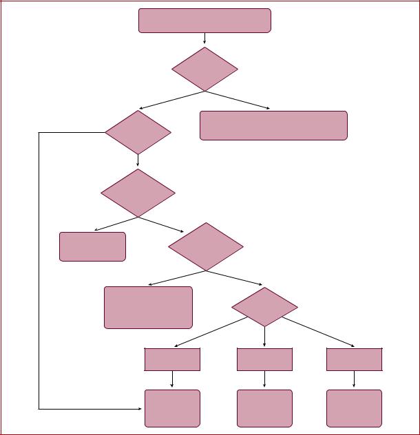
The regularity of the RR interval should be assessed (Figure 1). Irregular tachycardias may represent focal or multifocal AT, focal AF, and atrial flutter with varying AV conduction. Patterns of irregularity can sometimes be found, such as in atrial flutter conducted with Wenckebach periodicity. Irregular arrhythmias, such as multifocal AT, typically display variable P-wave morphologies, and varying PP, RR, and PR intervals. Atrial flutter can have fixed AV conduction and present as a regular tachycardia, and even AF may appear almost regular when very fast. Re-entrant tachycardias, whether micro or macro-re-entries, are usually regular. Incessant tachycardias may be the so-called permanent junctional reciprocating tachycardia (PJRT), focal AT, or, rarely, atypical AVNRT. Cycle length (CL) alternans (also called RR alternans) may be seen in AVNRT, but these changes are <15% of the tachycardia CL.47 If the irregularity exceeds 15% of the CL, a focal arrhythmia is much more likely.48 QRS alternans is a rare phenomenon in slow SVTs, which may not be related to CL alternans, and has been initially described with AVRT.49,50 However, this may be seen in any fast SVT.51
A change in ventricular CL preceded by a change in atrial CL is seen in AT or atypical AVNRT. A change in ventricular CL preceding a change in subsequent atrial CL favours typical AVNRT or AVRT.47,52 A fixed ventriculoatrial (VA) interval in the presence of variable RR intervals excludes AT.45
9.1.1.3 P/QRS relationship
According to their P/QRS relationships, SVTs are classified as having short or long RP intervals. Short-RP SVTs are those with RP intervals
2019 September 13 on guest by 1093/eurheartj/ehz467/5556821.abstract/doi/10-article-com/eurheartj/advance.oup.https://academic from Downloaded
12 |
ESC Guidelines |
|
|
|
Narrow QRS tachycardia |
|
|
|
|
(QRS≤120 ms) |
|
|
|
|
|
Regular |
|
|
|
|
tachycardia |
|
|
|
Yes |
No |
|
|
|
|
AF |
|
|
No |
Visible |
Focal AT or flutter with variable AV conduction |
|
|
Multifocal AT |
|
|
||
|
P waves? |
|
|
|
|
|
|
|
|
|
Yes |
|
|
|
|
Atrial rate |
|
|
|
|
greater than |
|
|
|
|
ventricular rate? |
|
|
|
Yes |
No |
|
|
|
Atrial flutter |
Ventricular rate |
|
|
|
Focal AT |
|
|
||
|
greater than |
|
|
|
AVNRT (rare) |
|
|
|
|
|
atrial rate |
|
|
|
|
|
|
|
|
|
Yes |
No |
|
|
|
High septal VT |
|
|
|
|
JET |
Consider |
|
|
|
AVNRT (rare) |
|
|
|
|
RP Interval |
|
|
|
Nodoventricular/fascicular-nodal |
|
|
||
|
|
|
||
|
re-entry (rare) |
|
|
|
|
Short (RP<PR) |
Short (RP<PR) |
Long (RP≥PR) |
|
|
RP≤90 ms |
RP>90 ms |
|
|
|
|
|
||
|
Typical AVNRT |
AVRT |
Focal AT |
2019 |
|
Focal AT |
|||
|
Atypical AVNRT |
AVRT |
||
|
JET |
|||
|
Focal AT |
Atypical AVNRT |
||
|
AVRT (rare) |
©ESC |
||
|
|
|
||
|
|
|
|
|
Figure 1 Differential diagnosis of narrow QRS tachycardia. Recording of a retrograde P wave should be sought by obtaining a 12 lead Electrocardiogram and, if necessary, using the Lewis leads or even an oesophageal lead connected to a pre-cordial lead (V1) with use of alligator clamps. The 90 ms cut-off is a rather arbitrary number used for surface electrocardiogram measurements if P waves are visible and is based on limited data. In the electrophysiology laboratory, the cut-off of the ventriculoatrial interval is 70 ms. Junctional ectopic tachycardia may also present with atrioventricular dissociation.
AF = atrial fibrillation; AT = atrial tachycardia; AV = atrioventricular; AVNRT = atrioventricular nodal re-entrant tachycardia; AVRT = atrioventricular reentrant tachycardia; JET = junctional ectopic tachycardia; RP = RP interval; VT = ventricular tachycardia.
2019 September 13 on guest by 1093/eurheartj/ehz467/5556821.abstract/doi/10-article-com/eurheartj/advance.oup.https://academic from Downloaded
ESC Guidelines |
13 |
|
|
shorter than one-half the tachycardia RR interval, whereas long-RP SVTs display RP>PR (Figure 1). Rarely, recording of U waves during
typical AVNRT may simulate a long-RP tachycardia.53
On EPS, a very short VA interval (<70 ms) usually indicates typical AVNRT, or less commonly focal AT, but has also been reported in AVRT.54 For surface ECG measurements, a cut-off interval of 90 ms has been shown to be useful and can be used if P waves are visible,55 but data on actual RP measurements during various types of SVT are scarce.
P waves similar to those in normal sinus rhythm suggest appropriate or inappropriate sinus nodal tachycardia, sinus nodal re-entrant tachycardia, or focal AT arising close to the sinus node. P waves different from those in sinus rhythm, and conducted with a PR interval equal to or longer than the PR in sinus rhythm, are typically seen in focal AT. In AT, the conduction to the ventricles may be fast (1:1) or slow (3:1 or 4:1). The possibility of atrial flutter with 2:1 conduction should also be considered if the ventricular rate during SVT is 150 b.p.m., as the atrial activity is usually 250 - 330 b.p.m. In the presence of antiarrhythmic medication in this setting, lowering the atrial rate may result in a higher ventricular rate in the absence of AV nodal blockade.
In the case of relatively delayed retrograde conduction that allows the identification of retrograde P waves, a pseudo r deflection in lead V1 and a pseudo S wave in the inferior leads are more common in typical AVNRT than in AVRT or AT.56,57 These criteria are specific (91 100%) but modestly sensitive (58 and 14%, respectively).56 A difference in RP intervals in leads V1 and III >20 ms is also indicative of AVNRT rather than AVRT due to a posteroseptal pathway.57 The presence of a QRS notch in lead aVL has also been found to be a reliable criterion suggesting AVNRT,58 while a pseudo r in aVR has been shown to have greater sensitivity and specificity than a pseudo r in V1 for the diagnosis of typical AVNRT.59 However, in all referenced studies, cases of AT or atypical AVNRT were limited or entirely absent.
AV block or dissociation during narrow QRS tachycardia is not often seen, but it rules out AVRT as both atria and ventricles are parts of the circuit. The development of bundle branch block (BBB) during SVT may also be helpful in the diagnosis of AVRT. BBB ipsilateral to the AP can result in CL prolongation due to VA prolongation, as the ventricular arm of the circuit is prolonged by conduction through the interventricular septum from the conducting bundle branch.60 However, it should be noted that lengthening of the VA interval may not necessarily result in CL prolongation, due to a potential switch of antegrade conduction from the slow to the fast AV nodal pathway.
9.1.2 Vagal manoeuvres and adenosine
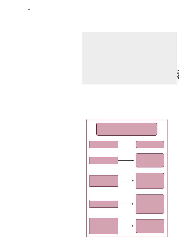
Vagal manoeuvres (such as carotid sinus massage) and adenosine injection may help in clinical diagnosis, particularly in situations in which the ECG during tachycardia is unclear. Possible responses to vagal manoeuvres and adenosine are shown in Table 8 and Figure 2.
Termination of the arrhythmia with a P wave after the last QRS complex is very unlikely in AT, and most common in AVRT and typical AVNRT. Termination with a QRS complex is often seen in AT, and possibly in atypical AVNRT. Adenosine does not interrupt mac- ro-re-entrant ATs (MRATs).61 Fascicular VTs, in particular, are
.
. verapamilbut not adenosine-sensitive. Most VTs, as opposed to
.
.
. SVTs, do not respond to carotid sinus massage, but a narrow QRS
.
.
. VT originating at the left bundle branch and terminated with carotid
.
.
. 62
. sinus massage has been reported.
.
.
.
Table 8 Possible responses of narrow QRS tachycardia to vagal manoeuvres and adenosine
(1)Slowing of AVN conduction and induction of intermittent AV block. Atrial electrical activity can thus be unmasked, revealing dissociated P waves (focal AT, atrial flutter, or AF waves).
(2)Temporary decrease in the atrial rate of automatic tachycardias (focal AT, sinus tachycardia, and JET).
(3)Tachycardia termination. This can happen by interrupting the re-entry circuit in AVNRT and AVRT by acting on the AVN that is part of the circuit. More rarely, sinus nodal re-entry and ATs due to triggered activity can slow down and terminate.
(4)No effect is observed in some cases.
AF = atrial fibrillation; AT = atrial tachycardia; AV = atrioventricular; AVN = atrioventricular node; AVNRT = atrioventricular nodal re-entrant tachycardia; AVRT = atrioventricular re-entrant tachycardia; JET = junctional ectopic tachycardia.
|
Adenosine |
|
|
in regular, narrow QRS tachycardia |
|
||
Response |
Diagnosis |
|
|
|
Inadequate dose/ |
|
|
No effect |
delivery |
|
|
|
High septal VT |
|
|
|
Sinus tachycardia |
|
|
Gradual slowing |
Automatic focal AT |
|
|
then reacceleration |
Junctional ectopic |
|
|
|
tachycardia |
|
|
|
AVNRT |
|
|
|
AVRT |
|
|
Sudden termination |
Sinus nodal re-entry |
|
|
|
Triggered focal AT |
|
|
|
(DADs) |
|
|
Persisting atrial |
Atrial flutter |
2019 |
|
tachycardia with |
|||
Micro-re-entrant |
|||
transient high-grade |
|||
focal AT |
|||
©ESC |
|||
AV block |
|||
|
|||
|
|
||
Figure 2 Responses of narrow-complex tachycardias to adenosine. AT = atrial tachycardia; AV = atrioventricular; AVNRT = atrioventricular nodal re-entrant tachycardia; AVRT = atrioventricular re-entrant tachycardia; DADs = delayed after-depolarizations; VT = ventricular tachycardia.
2019 September 13 on guest by 1093/eurheartj/ehz467/5556821.abstract/doi/10-article-com/eurheartj/advance.oup.https://academic from Downloaded

14 |
ESC Guidelines |
|
|
9.1.3 Electrophysiology study
Several electrophysiological techniques and manoeuvres can be employed in the electrophysiology laboratory for differential diagnosis of regular narrow QRS tachycardia.45 A detailed discussion is beyond the scope of these Guidelines.
9.2 Wide QRS (>120 ms) tachycardias
Wide QRS tachycardias can be VT, SVT conducting with BBB aberration, or antegrade conduction over an AP, with reported proportions of 80, 15, and 5%, respectively.63 The correct diagnosis of a VT is critical to management, as misdiagnosis and administration of drugs usually utilized for SVT can be harmful for patients in VT.64 Therefore, the default diagnosis should be VT until proven otherwise. The differential diagnosis includes:65
(1)SVT with BBB. This may arise due to pre-existing BBB or the development of aberrancy during tachycardia (so-called phase 3 block), which is more commonly, although not invariably, a right BBB (RBBB) pattern due to the longer RP of the right bundle branch.
(2)SVT with antegrade conduction over an AP, ‘pre-excited SVT’, which participates in the circuit (antidromic AVRT) or is a bystander during AF, focal AT/atrial flutter, or AVNRT.
(3)SVT with widening of QRS interval induced by drugs or electrolyte disturbances. Class IA and IC drugs cause use-dependent slowing of conduction, and class III drugs prolong refractoriness at His Purkinje tissue more than in the ventricular myocardium. They can both result in atypical BBB morphologies during SVT that mimics VT.
(4)Pacemaker-related endless loop tachycardia and artefacts, which can also mimic VT.
Table 9 Summary of key electrocardiographic criteria that suggest ventricular tachycardia rather than supraventricular tachycardia in wide complex tachycardia
AV dissociation |
Ventricular rate > atrial rate |
Fusion/capture beats |
Different QRS morphology from that |
|
of tachycardia |
Chest lead negative |
All precordial chest leads |
concordance |
negative |
RS in precordial leads |
- Absence of RS in precordial leads |
|
- RS >100 ms in any leada |
QRS complex in aVR |
• Initial R wave |
|
• Initial R or Q wave >40 ms |
|
• Presence of a notch of a predomi- |
|
nantly negative complex |
QRS axis 290 to 6180 |
Both in the presence of RBBB and |
|
LBBB morphology |
R wave peak time |
R wave peak time >50 ms |
in lead II |
|
RBBB morphology |
Lead V1: Monophasic R, Rsr’, biphasic |
|
qR complex, broad R (>40 ms), and a |
|
double-peaked R wave with the left |
|
peak taller than the right (the so-called |
|
‘rabbit ear’ sign) |
|
Lead V6: R:S ratio <1 (rS, QS patterns) |
LBBB morphology |
Lead V1: Broad R wave, slurred or |
|
notched-down stroke of the S wave, |
|
and delayed nadir of S wave |
|
Lead V6: Q or QS wave |
AV = atrioventricular; LBBB = left bundle branch block; RBBB = right bundle branch block.
aRS: beginning of R to deepest part of S.
9.2.1 Electrocardiographic differential diagnosis
If the 12 lead ECG is available in sinus rhythm, it can provide useful diagnostic information. Specifically, if the QRS morphology is identical during sinus rhythm and tachycardia, the arrhythmia is most likely not a VT. However, bundle branch re-entrant VTs and high septal VTs exiting close to the conduction system can have similar morphologies to sinus rhythm. The presence of a contralateral BBB pattern in sinus rhythm is more indicative of VT.
9.2.1.1 Atrioventricular dissociation
The presence of either AV dissociation or capture/fusion beats in the 12 lead ECG during tachycardia are key diagnostic features of VT. AV dissociation may be difficult to recognize because P waves are often hidden by wide QRS and T waves during a wide QRS tachycardia. P waves are usually more prominent in inferior leads and in modified chest lead placement (Lewis lead).63
The relationship between atrial and ventricular events is 1:1 or greater (more atrial than ventricular beats) in most SVTs (Table 9). AVNRT can be associated with 2:1 conduction,66 but this is rare. Although VA conduction can be found in <50% of patients with VT and a 1:1 relationship is possible, most VTs have a relationship <1:1 (more QRS complexes than P waves).
.
.
.
.
.
.
.
.
.
.
.
.
.
.
.
.
.
.
.
.
.
.
.
.
.
.
.
.
.
.
.
.
.
.
.
.
.
.
.
.
.
.
.
.
.
.
.
.
.
.
.
.
.
.
.
.
.
.
.
.
.
9.2.1.2 QRS duration
A QRS duration >140 ms with RBBB or >160 ms with left BBB (LBBB) pattern suggests VT. These criteria are not helpful for differentiating VT from SVT in specific settings, such as pre-excited SVT, or when class IC or class IA antiarrhythmic drugs are administered.67
9.2.1.3 QRS axis
As VT circuits (especially post-MI or in cardiomyopathies) frequently lie outside the normal His Purkinje network, significant axis shifts are likely to occur, enabling diagnosis. Therefore, in patients with SVT and aberrancy, the QRS axis is confined between -60 and þ120 . In particular, extreme axis deviation (axis from -90 to ±180 ) is strongly indicative of VT, both in the presence of RBBB and LBBB.65
9.2.1.4 Chest lead concordance
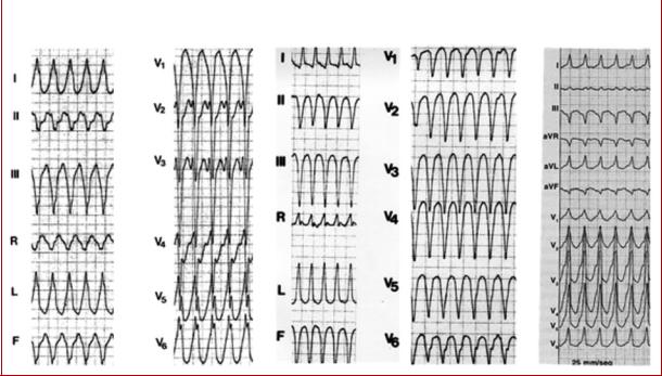
The presence of negative chest lead concordance (all QRS complexes negative V1 V6) (Figure 3) is almost diagnostic of VT, with a specificity >90%, but is only present in 20% of VTs. Positive concordance can be indicative of VT or an antidromic tachycardia utilizing a left posterior or left lateral AP.68
2019 September 13 on guest by 1093/eurheartj/ehz467/5556821.abstract/doi/10-article-com/eurheartj/advance.oup.https://academic from Downloaded
ESC Guidelines |
|
15 |
||
|
|
|
|
|
|
|
|
|
|
|
Antidromic AVRT |
VT with negative |
VT with positive |
|
|
|
concordance |
concordance |
|
©ESC 2019
Figure 3 Examples of positive and negative chest lead concordance in VT.
AVRT = atrioventricular re-entrant tachycardia; VT = ventricular tachycardia.
9.2.1.5 Right bundle branch block morphology
Lead V1: Typical RBBB aberrancy has a small initial r’, because in RBBB the high septum is activated primarily from the left septal bundle. Therefore, the following patterns are evident: rSR0, rSr0, or rR0 in lead V1. However, in VT, the activation wavefront progresses from the left ventricle (LV) to the right precordial lead V1, in a way that a prominent R wave (monophasic R, Rsr0, biphasic qR complex, or broad R >40 ms) will be more commonly seen in lead V1. Additionally, a double-peaked R wave (M pattern) in lead V1 favours VT if the left peak is taller than the right peak (the so-called ‘rabbit ear’ sign). A taller right rabbit ear characterizes the RBBB aberrancy but does not exclude VT.
Lead V6: A small amount of normal right ventricular voltage is directed away from lead V6. As this is a small vector in RBBB aberrancy, the R:S ratio is >1. In VT, all of the right ventricular voltage, and some of the left, is directed away from V6, leading to an R:S ratio <1 (rS and QS patterns). An RBBB morphology with an R:S ratio in V6 of <1 is seen rarely in SVT with aberrancy, mainly when the patient has a left axis deviation during sinus rhythm.
Differentiating fascicular VT from SVT with bifascicular block (RBBB and left anterior hemiblock) is very challenging. Features that indicate SVT in this context include QRS >140 ms, r’ in V1, overall negative QRS in aVR, and an R/S ratio >1 in V6.44
9.2.1.6 Left bundle branch block morphology
Lead V1: As stated above for RBBB, for the same reasons, the presence of a broad R wave, slurred or notched-down stroke of
.
.
.
.
.
.
.
.
.
.
.
.
.
.
.
.
.
.
.
.
.
.
.
.
.
.
.
.
.
.
.
.
.
.
.
.
.
.
.
.
.
.
.
.
.
.
.
.
.
.
.
.
.
.
.
.
.
.
.
.
.
.
.
.
.
.
.
.
.
.
.
.
.
.
.
.
.
.
.
.
.
.
the S wave, and delayed nadir of the S wave are strong predictors of VT.
Lead V6: In true LBBB, no Q wave is present in the lateral precordial leads. Therefore, the presence of any Q or QS wave in lead V6 favours VT, indicating that the activation wavefront is moving away from the LV apical site.
These morphology criteria are not fulfilled in any lead in 4% of SVTs and 6% of VTs, and in one-third of cases when one lead (V1 or V6) favours one diagnosis, the other favours the opposite diagnosis (VT in one lead and SVT in the other, and vice versa).69,70
A number of algorithms have been developed to differentiate VT from SVT.69,71,72 Detailed presentation and comments are beyond the scope of these Guidelines, and can be found in the 2018 European Heart Rhythm Association/Heart Rhythm Society/Asia Pacific Heart Rhythm Society/Sociedad Latinoamericana de Estimulacion Cardıaca y Electrofisiologıa consensus document.3
All of these criteria have limitations. Conditions such as bundle branch re-entrant tachycardia, fascicular VT, VT with exit site close to the His Purkinje system, and wide QRS tachycardia occurring during antiarrhythmic drug treatment are difficult to diagnose using the mentioned morphological criteria. Differentiating VT from antidromic AVRT is extremely difficult for the very fact that the QRS morphology in antidromic AVRT is similar to that of a VT, with its origin at the insertion of the AP in the ventricular myocardium. An algorithm has been derived for differential diagnosis, based on the analysis of 267 wide QRS tachycardias, consisting of VT and antidromic
2019 September 13 on guest by 1093/eurheartj/ehz467/5556821.abstract/doi/10-article-com/eurheartj/advance.oup.https://academic from Downloaded