Section Ill • Gross Anatomy
Clinical Correlate
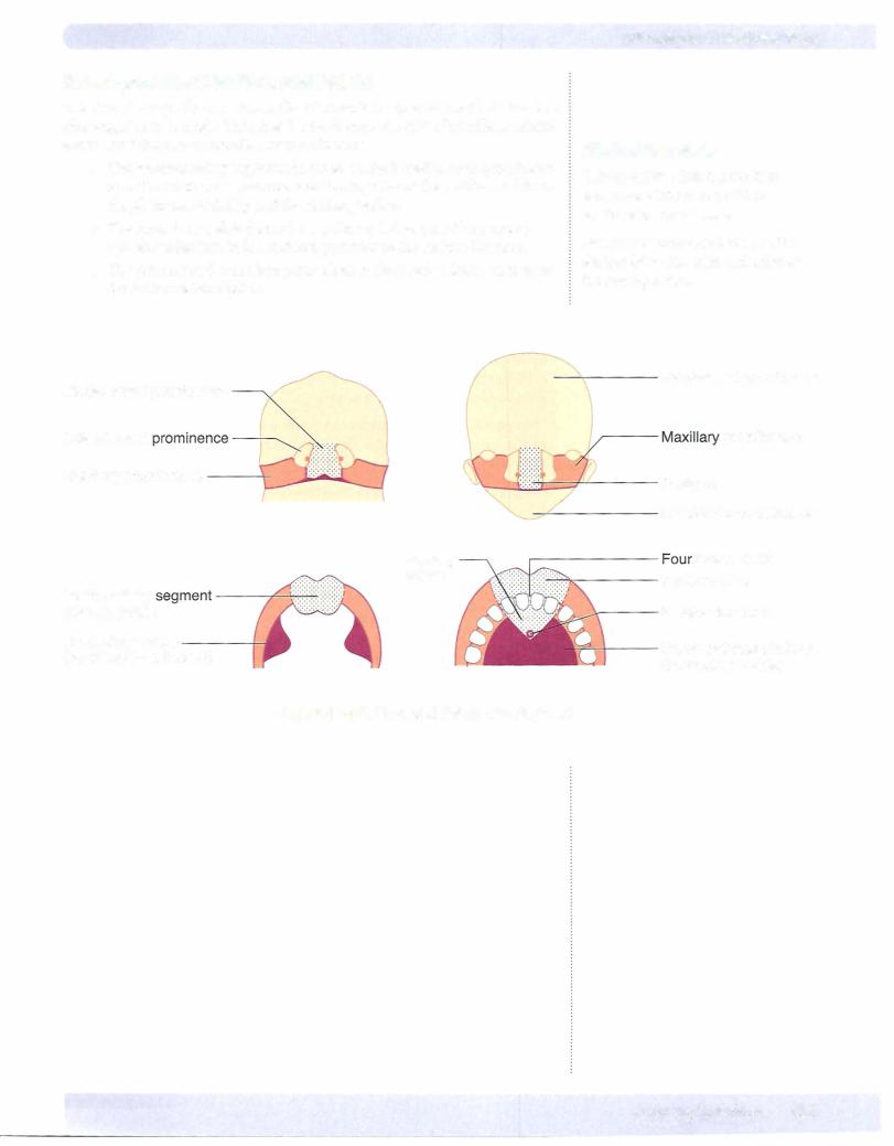
Robin sequence presents with a triad of poor mandibular growth, cleft palate, and a posteriorly placed tongue.
Treacher Collins syndrome also presents with mandibular hypoplasia, zygomatic hypoplasia, down-slanted palpebral fissures, colobomas, and malformed ears.
Clinical Correlate
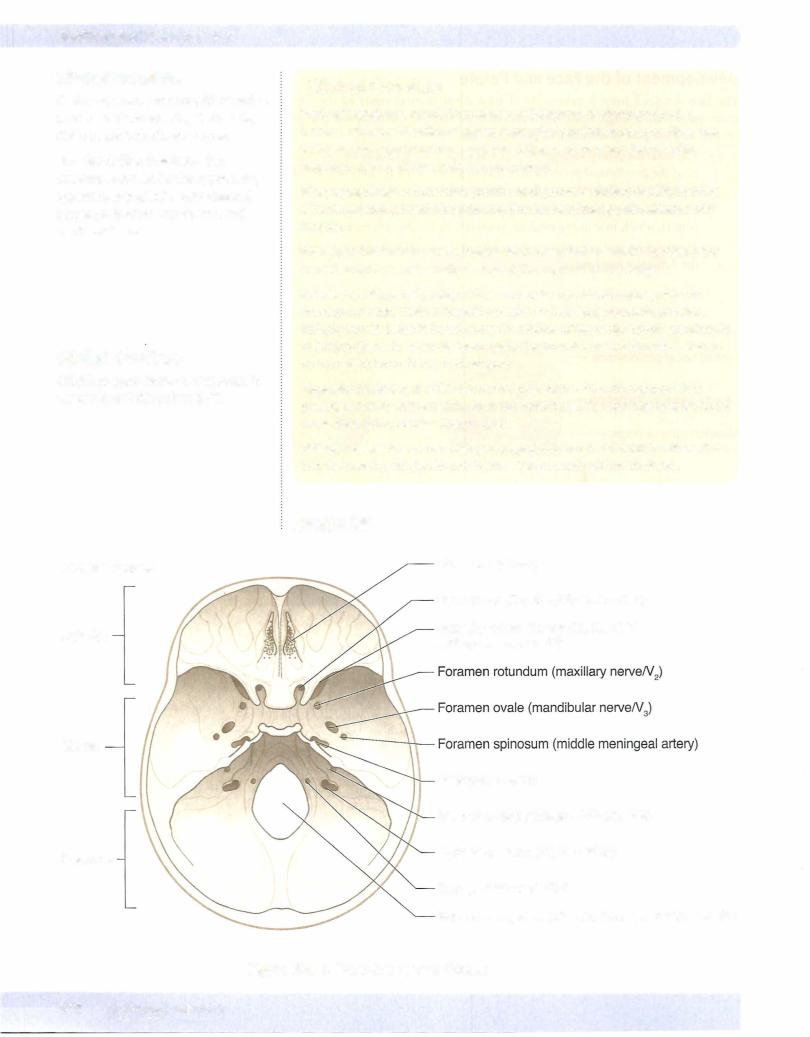
Cribriform plate fractures may result in dysosmia and rhinorrhea (CSF).
Clinical Correlate
First arch syndrome results from abnormal formation of pharyngeal arch 1 because of faulty migration of neural crest cells, causing facial anomalies. Two well-described syndromes are Treacher Collins syndrome and Pierre Robin sequence. Both defects involve neural crest cells.
Pharyngeal fistula occurs when pouch 2 and groove 2 persist, thereby forming a fistula generally found along the anterior border of the sternocleidomastoid muscle.
Pharyngeal cyst occurs when pharyngeal grooves that are normally obliterated persist, forming a cyst usually located at the angle of the mandible.
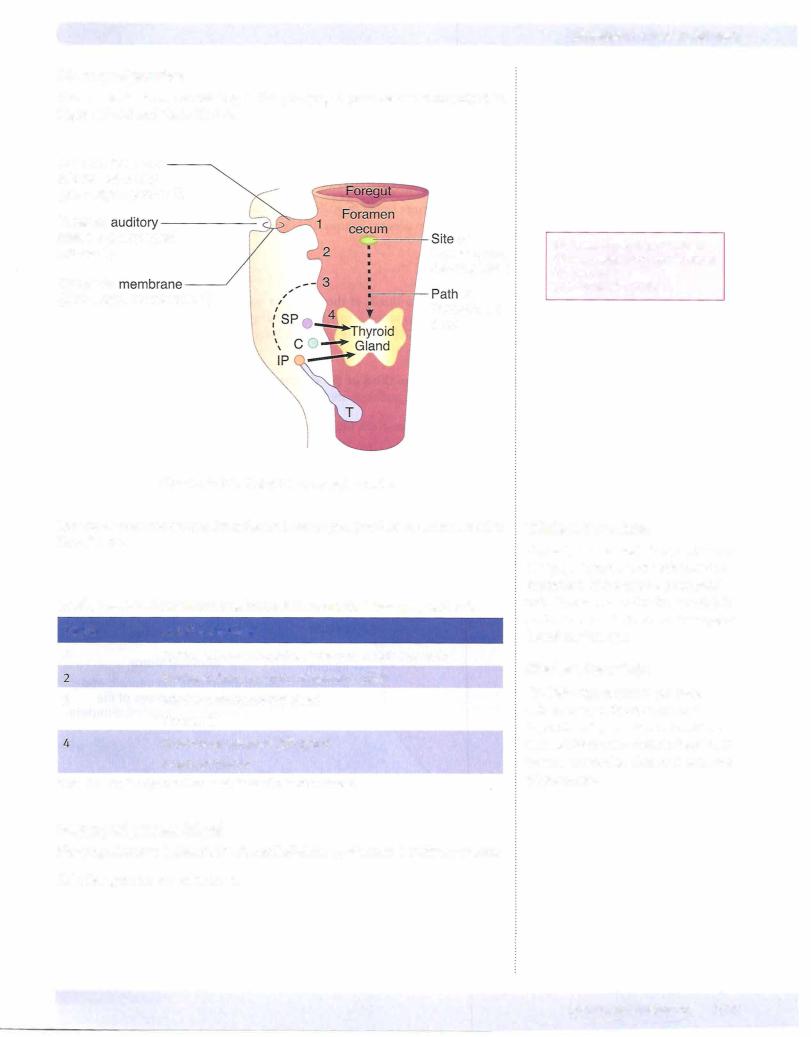
Ectopic thyroid, parathyroid, or thymus results from abnormal migration of these glands from their embryonic position to their adult anatomic position. Ectopic thyroid tissue is found along the midline ofthe neck. Ectopic parathyroid or thymus tissue is generally found along the lateral aspect ofthe neck. May be an important issue during neck surgery.
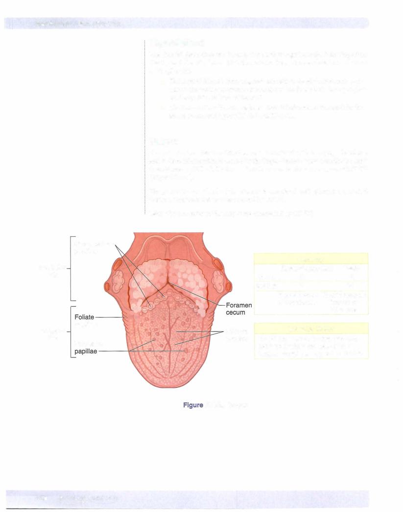
Thyroglossal duct cyst or fistula occurs when parts of the thyroglossal duct persist, generally in the midline near the hyoid bone. The cyst may also be found at the base ofthe tongue (lingual cyst).
DiGeorge sequence occurs when pharyngeal pouches 3 and 4 fail to differentiate into the parathyroid glands and thymus. Neural crest cells are involved.
CRANIUM