Материал: Kaplan USMLE-1 (2013) - Anatomy
Chapter 6 • Head and Neck
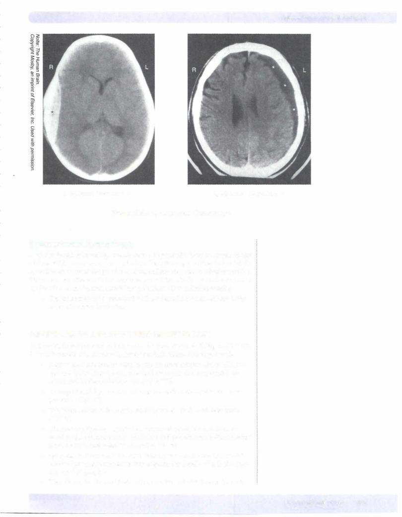
A. Epidural Hematoma* |
B. Subdural Hematoma* |
Figure 111-6-14. lntracranial Hemorrhage
Subarachnoid Hemorrhage
A subarachnoid hemorrhage results from a rupture of a berry aneurysm in the circle ofWillis. The most common site is in the anterior part ofthe circle ofWillis at the branch point ofthe anterior cerebral and anterior communicating arteries. Other common sites are in the proximal part of the middle cerebral artery or at the junction ofthe internal carotid and posterior communicating arteries.
•Typical presentation associated with a subarachnoid hemorrhage is the onset of a severe headache.
ORBITAL MUSCLES AND THEIR INNERVATION
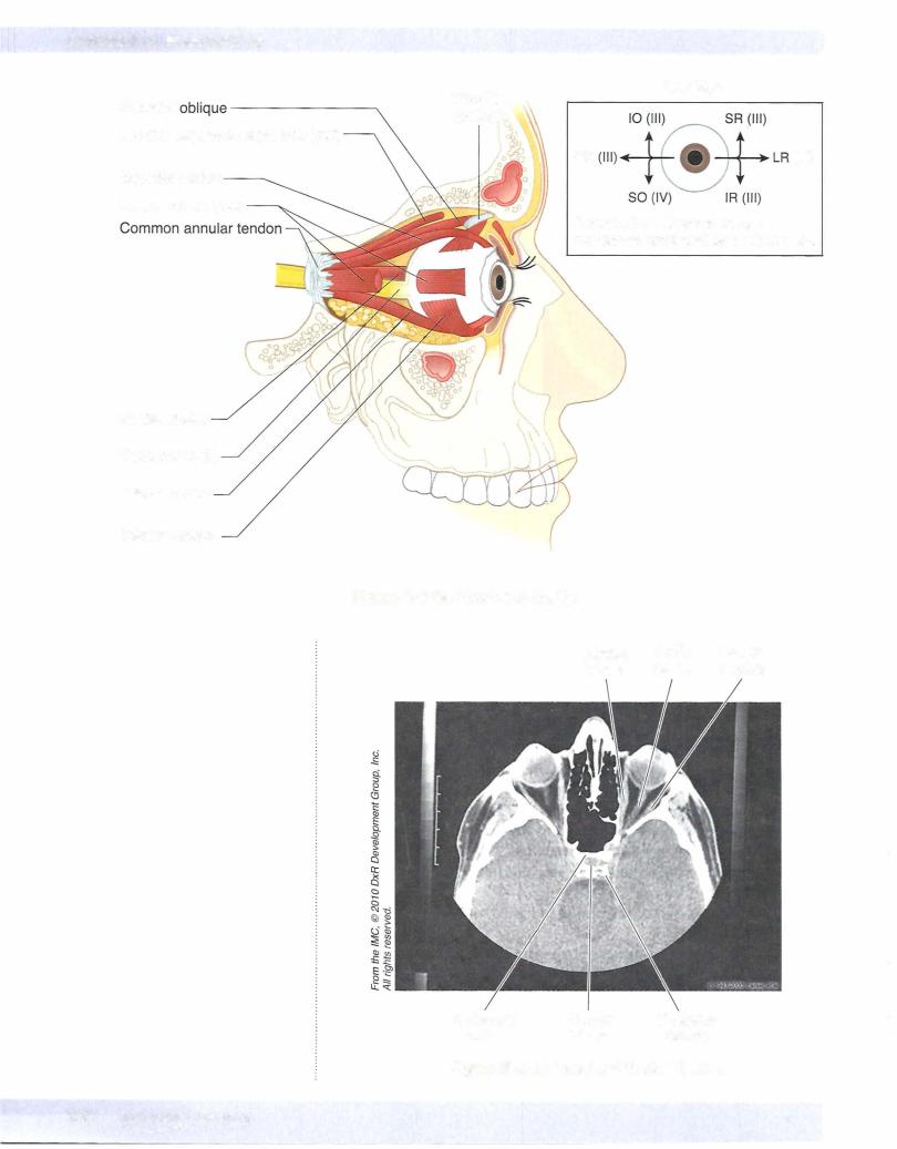
In the orbit, thereare 6 ex.traocular muscles thatmovethe eyeball (Figure III-6-16).
A seventh muscle, the levator palpebrae superioris, elevates the upper eyelid.
•Pour ofthe 6 ex.traocular muscles (the superior, inferior, and medial rec tus, and the inferior oblique, plus the levatorpalpebrae superioris) are innervated by the oculomotor nerve (CN III).
•The superior oblique muscle is the only muscle innervated by the troch lear nerve (CN IV).
•The lateral rectus is the only muscle innervated by the abducens nerve (CN VI).
•The levator palpebrae superioris is composed ofskeletal muscle inner vated by the oculomotor nerve (CN III) and smooth muscle (the superior tarsal muscle) innervatedby sympathetic fibers.
•Sympathetic fibersreachthe orbit from a plexus on the internal carotid artery ofpostganglionic axons that originate from cell bodies in the supe rior cervical ganglion.
•The orbital muscles and their actions are illustrated in Figure III-6- 1 5.
MEDICAL 327

SECTION
Neuroscience