Материал: 2019 ESC - supraventricular tachycardia

ESC Guidelines |
21 |
monitoring. The evaluation of a patient suspected of having POTS should eliminate other causes of sinus tachycardia such as hypovolaemia, anaemia, hyperthyroidism, pulmonary embolus, or pheochromocytoma.178 The clinical history should focus on defining the chronicity of the condition, possible causes of orthostatic tachycardia, modifying factors, impact on daily activities, and potential triggers.
11.1.1.4.2 Therapy. Non-pharmacological treatments should be attempted first in all patients. These include withdrawing medications that might worsen POTS, such as norepinephrine transport inhibitors, increasing blood volume with enhanced salt and fluid intake, reducing venous pooling with compression garments, and limiting deconditioning. Patients should engage in a regular, graduated, and supervised exercise programme featuring aerobic reconditioning with some resistance training for the thighs. Initially, exercise should be restricted to non-upright exercise, including the use of rowing machines and swimming, to minimize orthostatic stress on the heart.180 182
If non-pharmacological approaches prove ineffective, pharmacological therapies may be targeted at specific aspects. Patients strongly suspected of having hypovolaemia should drink >2 3 L of water per day, and dietary salt intake should be increased to 10 12 g/day if tolerated. Midodrine significantly reduces orthostatic tachycardia but to a lesser degree than i.v. saline.170 Midodrine has a rapid onset with only brief effects and is usually administered three times daily. The drug should only be administered during daytime hours as it can cause supine hypertension. To reduce unpleasant sinus tachycardia and palpitations, low-dose propranolol [10 20 mg per os (p.o.)] acutely lowers standing heart rate and improves symptoms in patients with POTS, while higher doses of propranolol are less well tolerated.172 Long-acting propranolol does not improve the quality of life of patients with POTS.167 Non-selective beta-blockers are preferable because they additionally block epinephrine-mediated beta-2-vasodilation, but other beta-blockers have not been adequately studied. Pyridostigmine, a cholinergic agonist that works by inhibiting acetylcholinesterase, can increase parasympathetic autonomic tone and has a lower risk of hypertension compared with other medications. Potential side effects include abdominal cramping, diarrhoea, and muscle cramps.173,174 Ivabradine slows sinus rates without affecting blood pressure, and in an open-label study 60% of patients with POTS had symptomatic improvement.175 Ivabradine should ideally be administered with concomitant beta-blockers for long-term therapy.151
11.1.2 Focal atrial tachycardia
Focal AT is defined as an organized atrial rhythm >100 b.p.m. initiated from a discrete origin and spreading over both atria in a centrifugal pattern. The ventricular rate varies, depending on AV nodal conduction. In asymptomatic young people (<50 years of age), the prevalence of focal AT has been reported to be as low as 0.34% with an increased prevalence of 0.46% in symptomatic arrhythmia patients.183 Most studies have reported no influence of sex.
Symptoms may include palpitations, shortness of breath, chest pain, and rarely syncope or presyncope. The arrhythmia may be
.
.
.
.
.
.
.
.
.
.
.
.
.
.
.
.
.
.
.
.
.
.
.
.
.
.
.
.
.
.
.
.
.
.
.
.
.
.
.
.
.
.
.
.
.
.
.
.
.
.
.
.
.
.
.
.
.
.
.
.
.
.
.
.
.
.
.
.
.
.
.
.
.
.
.
.
.
.
.
.
.
.
.
.
.
.
.
.
.
.
.
.
.
.
.
.
.
.
.
.
.
.
.
.
.
.
.
.
.
.
.
.
.
.
.
.
.
.
.
.
.
.
.
.
.
.
.
.
.
.
.
.
.
.
.
.
.
.
.
.
.
.
.
.
.
.
.
.
.
.
.
.
.
.
.
.
.
.
.
.
.
.
.
.
.
.
.
.
.
.
.
.
.
sustained or incessant. Dynamic forms with recurrent interruptions and reinitiations may be frequent.
In patients with PV-related AT, the focus is located at the ostium
of the vein (or within 1 cm of the designated ostium)184 rather than further distally (2 4 cm).185
11.1.2.1 Diagnosis
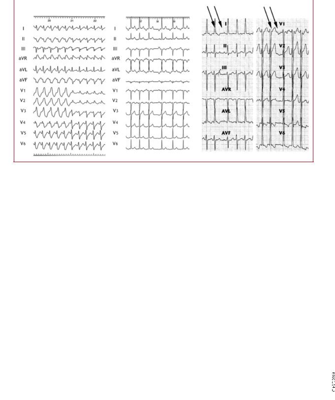
P wave identification from a 12 lead ECG recording during tachycardia is critical (Figure 7). Depending on the AV conduction and AT rate, the P waves may be hidden in the QRS or T waves. The P waves are monomorphic with stable CL, which helps to rule out organized AF. Adenosine injection can help by slowing the ventricular rate or, less frequently, by terminating focal AT. A discrete P wave with an intervening isoelectric interval suggests a focal AT. However, distinguishing focal from macro-re-entrant arrhythmias by surface ECG is not always possible. The presence of an isoelectric line does not rule out a macro-re-entrant mechanism, particularly in the presence of scar atrial tissue (from structural heart disease or previous extensive ablation/surgery procedures). In a normal heart and in the absence of previous ablation, the usual ECG localization rules apply,186 but their value in localizing the origin of the arrhythmia is also limited in this context. Focal AT may arise from any site in both atria, but particular sites of predilection in the normal heart rate are the crista terminalis, the tricuspid and mitral valve annulus, and within the thoracic veins joining the atria.46,186 A negative P wave in lead I and aVL suggests an LA origin. V1 is negative when the arrhythmia source or exit is in the lateral right atrium, while septal right atrial (RA) and LA origins show biphasic or positive P waves (Figure 7). Negative P waves in the inferior leads suggest a caudal origin, whereas positive P waves in those leads favour a superior location.
11.1.2.2 Acute therapy
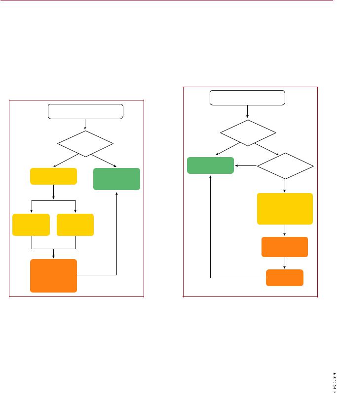
Hard data for an evidence-based choice of drugs for the acute therapy of focal AT are scarce. In general, acute therapy may be initiated with beta-blockers or calcium channel blockers, which may terminate focal ATs or slow the ventricular rate (Figure 8).92,94,187,188 Adenosine (i.v.) may terminate AT [delayed after-depolarizations (DAD)-triggered AT], but the tachycardia may also continue with AV block. Class IA, IC, and III drugs may also be effective, by prolonging refractoriness or suppressing automaticity.189 191 Amiodarone may also be used for cardioversion or slowing of the ventricular rate,192 but the efficacy of rate control is unproven in critically ill patients with atrial arrhythmias.193 DC cardioversion is usually effective in acutely terminating the tachycardia, irrespective of the mechanism. However, in incessant forms of focal AT due to enhanced automaticity, the arrhythmia reinitiates, and repeating DC cardioversion is unlikely to be appropriate.
11.1.2.3 Catheter ablation
Catheter ablation is the treatment of choice for recurrent focal AT, especially for incessant AT due to which TCM ensues (Figure 9).196 Distinguishing macro-re-entrant from focal ATs is critical for the ablation strategy. Focal ATs, as well as localized/ micro-re-entry ATs, display a centrifugal activation pattern that spreads throughout the atria. Mapping and ablation of focal ATs is based on determining the earliest activation site. In PV-related AT, focal ablation may be performed, but electrical isolation of both
2019 September 13 on guest by 1093/eurheartj/ehz467/5556821.abstract/doi/10-article-com/eurheartj/advance.oup.https://academic from Downloaded
22 |
|
|
ESC Guidelines |
|
|
|
|
|
|
|
|
|
|
|
|
A |
B |
C |
|
©ESC 2019
Figure 7 Focal atrial tachycardia. (A) Focal atrial tachycardia originating at the lateral right atrium conducted initially with full and then incomplete right branch bundle block aberration. (B) Focal atrial tachycardia originating at the left atrium (left superior pulmonary vein). (C) Focal atrial tachycardia from the right atrial appendage. Atrioventricular dissociation during carotid sinus massage (P waves indicated by arrows).
Recommendations for the therapy of focal atrial tachycardia
|
Recommendation |
Classa |
Levelb |
|
|
Acute therapy |
|
|
|
|
Haemodynamically unstable patients |
|
|
|
|
Synchronized DC cardioversion is recommended for haemodynamically unstable patients.86 88 |
I |
B |
|
|
Haemodynamically stable patients |
|
|
|
|
Adenosine (6 18 mg i.v. bolus) should be considered.92 94 |
IIa |
B |
|
|
Beta-blockers (i.v. esmolol or metoprolol) should be considered in the absence of decompensated HF, if adenosine fails.187,188 |
IIa |
C |
|
|
Verapamil or diltiazem (i.v.) should be considered for haemodynamically stable patients in the absence of hypotension or HFrEF, |
IIa |
C |
|
|
if adenosine fails.92,94 |
|
||
|
|
|
|
|
|
If the above measures fail, the following may be used: |
|
|
|
|
• i.v. ibutilide191; |
IIb |
C |
|
|
• or i.v. flecainide or propafenone189,190; |
|
||
|
|
|
|
|
|
• or i.v. amiodarone.192 |
|
|
|
|
Synchronized DC cardioversion is recommended when drug therapy fails to convert or control the tachycardia.87,88 |
I |
B |
|
|
Chronic therapy |
|
|
|
|
Catheter ablation is recommended for recurrent focal AT, especially if incessant or causing TCM.184,187,194 197 |
I |
B |
|
|
Beta-blockers or non-dihydropyridine calcium channel blockers (verapamil or diltiazem in the absence of HFrEF), or propafenone or fle- |
IIa |
C |
|
|
cainide in the absence of structural or ischaemic heart disease, should be considered if ablation is not desirable or feasible.188 190,198 |
|
||
|
|
|
|
|
|
Ivabradine with a beta-blocker may be considered if the above measures fail.199,200 |
IIb |
C |
|
|
Amiodarone may be considered if the above measures fail.201,202 |
IIb |
C |
|
i.v. verapamil and diltiazem are contraindicated in the presence of hypotension or HFrEF. i.v. beta-blockers are contraindicated in the presence of decompensated heart failure. i.v. ibutilide is contraindicated in patients with prolonged QTc interval.
i.v. flecainide and propafenone are contraindicated in patients with ischaemic or structural heart disease. They also prolong the QTc interval but much less than class III agents. i.v. amiodarone prolongs the QTc but torsades des pointes is rare.
AT = atrial tachycardia; DC = direct current; HF = heart failure; HFrEF = heart failure with reduced ejection fraction; i.v. = intravenous; TCM = tachycardiomyopathy. aClass of recommendation.
bLevel of evidence.
2019 September 13 on guest by 1093/eurheartj/ehz467/5556821.abstract/doi/10-article-com/eurheartj/advance.oup.https://academic from Downloaded
ESC Guidelines |
23 |
the culprit PV along with other PVs may be preferred. Catheter ablation is reported to have a 75 100% success rate.184,187,194 197 Table 11 presents a synopsis of success rates and complications of
catheter ablation for the most common SVTs in the current
era.11 13,203 208
11.1.2.4 Chronic therapy
Studies addressing chronic therapy are also limited, and thus firm conclusions cannot be drawn. Beta-blockers and calcium channel
.
.
.
.
.
.
.
.
.
.
.
.
.
.
.
.
.
.
.
.
.
.
.
.
blockers may be effective, and there is a low risk of side effects (Figure 9).188 Class IC drugs may be effective if first-line therapy has failed.189,190,198 Ivabradine may also be effective in focal AT,199,200 and ideally should be given with a beta-blocker. Amiodarone has been tried in the young and paediatric populations,201,202 and theoretically should be attractive in patients with impaired LV function. However, its long-term efficacy is limited by its side effects.
|
|
|
|
|
Focal AT |
|
|
|
|
Focal AT |
|
|
|
|
|
|
|
|
|
|
Recurrent or |
|
|
|
|
Haemodynamic |
|
|
incessant |
|
|
|
|
|
Yes |
|
No |
|
|
|
|
instability |
|
|
|
||
|
|
|
|
|
|
|
|
|
No |
Yes |
|
|
|
|
|
|
|
|
|
Catheter ablation |
|
Drug therapy |
|
|
Adenosine |
Synchronized |
|
(I B) |
No |
desirable |
|
|
|
|
|
|
|
||
|
(IIa B) |
cardioversion |
|
|
|
|
|
|
|
(I B) |
|
|
|
Yes |
|
If ineffective |
If ineffective |
|
|
|
Beta-blocker or |
|
|
|
|
|
|
|
|
|
|
|
|
|
|
|
|
verapamil or diltiazem |
|
|
|
|
|
|
|
or propafenone |
|
i.v. verapamil |
|
i.v. |
|
|
|
or flecainide |
|
|
|
|
|
(IIa C) |
|
||
or diltiazem |
beta-blocker |
|
|
|
|
|
|
(IIa C) |
|
(IIa C) |
|
|
If ineffective |
|
|
|
If ineffective |
|
|
|
|
Ivabradine with a |
|
|
|
|
|
|
|
beta-blocker |
|
|
|
|
|
|
|
(IIb C) |
|
|
i.v. ibutilde |
If ineffective |
|
|
If ineffective |
|
|
or flecainide |
2019 |
|
|
Amiodarone |
2019 |
||
or propafenone |
|
|
|
||||
or amiodarone |
|
If ineffective |
(IIb C) |
||||
|
(IIb C) |
|
©ESC |
|
©ESC |
||
|
|
or not tolerated |
|
||||
|
|
|
|
|
|||
|
|
|
|
|
|
||
Figure 8 Acute therapy of focal atrial tachycardia. |
|
Figure 9 Chronic therapy of focal atrial tachycardia. |
|
||||
AT = atrial tachycardia; i.v. = intravenous. AT = atrial tachycardia.
Table 11 Average success and complication rates of catheter ablation for supraventricular tachycardia
|
Acute success (%) |
Recurrence (%) |
Complications (%) |
Mortality (%) |
Focal AT |
85 |
20 |
1.4a |
0.1 |
Cavotricuspid-dependent atrial flutter |
95 |
10 |
2b |
0.3 |
AVNRT |
97 |
2 |
0.3c |
0.01 |
AVRT |
92 |
8 |
1.5d |
0.1 |
Success rates, recurrence, and complications for focal atrial tachycardia and atrioventricular re-entrant tachycardia vary, being dependent on the location of the focus or pathway, respectively.11 13,203 208
aVascular complications, AV block, and pericardial effusion.
bVascular complications, stroke, myocardial infarction, and pericardial effusion. cVascular complications, AV block, and pericardial effusion.
dVascular complications, AV block, myocardial infarction, pulmonary thromboembolism, and pericardial effusion.
AT = atrial tachycardia; AV = atrioventricular; AVNRT = atrioventricular nodal re-entrant tachycardia; AVRT = atrioventricular re-entrant tachycardia.
2019 September 13 on guest by 1093/eurheartj/ehz467/5556821.abstract/doi/10-article-com/eurheartj/advance.oup.https://academic from Downloaded
24 |
ESC Guidelines |
11.1.3 Multifocal atrial tachycardia
Multifocal AT is defined as a rapid, irregular rhythm with at least three distinct morphologies of P waves on the surface ECG. Multifocal AT is commonly associated with underlying conditions, including pulmonary disease, pulmonary hypertension, coronary disease, and valvular heart disease, as well as hypomagnesaemia and theophylline therapy.209 It may also be seen in healthy infants under 1 year of age, and carries a good prognosis in the absence of underlying cardiac disease.210
It may be difficult to distinguish multifocal AT from AF on a single ECG trace, so a 12 lead ECG is indicated to confirm the diagnosis. On the ECG, the atrial rate is >100 b.p.m. Unlike AF, there is a distinct isoelectric period between visible P waves. The PP, PR, and RR intervals are variable. Although it is assumed that the variability of P-wave morphology implies a multifocal origin, there have been very few mapping studies of multifocal AT.
11.1.3.1 Therapy
The first-line treatment is management of the underlying condition. Magnesium (i.v.) may also be helpful in patients, even in those with normal magnesium levels.211 Antiarrhythmic medications, in general, are not helpful in suppressing multifocal AT.209 Management often involves slowing conduction at the AVN level to control heart rate. Verapamil has some efficacy in patients with multifocal AT who do not have ventricular dysfunction, sinus node dysfunction, or AV block.212,213 Metoprolol has been found to be superior to verapamil,214 and beta-blockers can be used with caution to treat multifocal AT in the absence of respiratory decompensation, sinus node
Recommendations for the therapy of multifocal atrial tachycardia
|
Recommendation |
Classa |
Levelb |
|
|
Acute therapy |
|
|
|
|
Treatment of an underlying condition is rec- |
I |
C |
|
|
ommended as a first step, if feasible.209 |
|
||
|
|
|
|
|
|
i.v. beta-blockers, or i.v. non-dihydropyridine |
|
|
|
|
calcium channel blockers (verapamil or diltia- |
IIa |
B |
|
|
zem) should be considered.213,214 |
|
|
|
|
Chronic therapy |
|
|
|
|
Oral verapamil or diltiazem should be consid- |
|
|
|
|
ered for patients with recurrent symptomatic |
IIa |
B |
|
|
multifocal AT in the absence of HFrEF.217,218 |
|
|
|
|
A selective beta-blocker should be considered |
|
|
|
|
for patients with recurrent symptomatic multi- |
IIa |
B |
|
|
focal AT.214,219 |
|
|
|
|
AV nodal ablation followed by pacing (prefera- |
|
|
|
|
ble biventricular or His-bundle pacing) should |
|
|
|
|
be considered for patients with LV dysfunction |
IIa |
C |
|
|
due to recurrent multifocal AT refractory to |
|
|
|
|
drug therapy.216 |
|
|
|
AT = atrial tachycardia; HF = heart failure; HFrEF = heart failure with reduced ejection fraction; i.v. = intravenous; LV = left ventricular.
i.v. verapamil and diltiazem are contraindicated in the presence of hypotension or HFrEF.
i.v. beta-blockers are contraindicated in the presence of decompensated heart failure.
aClass of recommendation. bLevel of evidence.
. |
|
|
|
|
|
. |
dysfunction, or AV block. There has also been a case report on the |
||||
. |
|||||
. |
215 |
|
|
||
. |
|
|
|||
. |
successful use of ibutilide. |
In symptomatic and medically refractory |
|||
. |
|
|
|
|
|
. |
cases in which LV deterioration is seen, AV nodal modification may |
||||
. |
|||||
. |
|
|
216 |
|
|
. |
|
|
|
|
|
. |
be used for the control of ventricular rate. |
|
|
||
. |
|
|
|
|
|
. |
|
|
|
|
|
. |
|
|
|
|
|
. |
|
|
|
|
|
. |
|
|
|
|
|
. |
|
|
|
|
|
. |
|
|
|
|
|
. |
11.1.4 Macro-re-entrant atrial tachycardias |
||||
. |
|||||
. |
|
|
|
|
|
. |
Atrial flutter and focal AT are traditionally defined according to the |
||||
. |
|||||
. |
|
|
|
|
|
. |
ECG appearance: continuous regular electrical activity, most commonly |
||||
. |
|||||
. |
|
|
|
|
|
. |
a saw-tooth pattern, vs. discrete P waves with an isoelectric line in |
||||
. |
|||||
. |
|
|
|
|
|
. |
between. ECGs with flutter-like appearances are mostly due to macro- |
||||
. |
|||||
. |
|
|
|
|
|
. |
re-entrant atrial circuits but micro-re-entry is also possible. However, |
||||
. |
|||||
. |
|
|
|
|
|
. |
MRATs with a significant part of the activation of the circuit in pro- |
||||
. |
|||||
. |
|
|
|
220 |
|
. |
|
|
|
||
. |
tected areas may display a focal AT pattern, with discrete P waves. |
||||
. |
|
|
|
|
|
. |
|
|
|
|
|
. |
|
|
|
|
|
. |
|
|
|
|
|
. |
|
|
|
|
|
. |
11.1.4.1 Cavotricuspid isthmus-dependent macro re-entrant AT |
||||
. |
|||||
. |
|
|
|
|
|
. |
|
|
|
|
|
. |
|
|
|
|
|
. |
|
|
|
|
|
. 11.1.4.1.1 Typical atrial flutter: common (counter-clockwise) and |
|||||
. |
reverse (clockwise). Typical common atrial flutter is the most fre- |
||||
. |
|||||
. |
|||||
. |
|
|
|
|
|
. |
quent cavotricuspid isthmus (CTI)-dependent flutter, i.e. a macro-re- |
||||
. |
|||||
. |
|
|
|
|
|
. |
entry circuit around the tricuspid annulus using the CTI as a critical |
||||
. |
|||||
. |
|
|
|
|
|
. |
passage at the inferior boundary. Activation goes downward in the RA |
||||
. |
|||||
. |
|
|
|
|
|
. |
free wall, through the CTI, and ascends in the right septum. Activation |
||||
. |
|||||
. |
|
|
|
|
|
. |
of the LA is passive. The upper part of the circuit may be anterior or |
||||
. |
|||||
. |
|
|
|
|
|
. |
|
|
|
|
|
. posterior to the superior vena cava. This activation is also known as |
|||||
. |
|
|
|
|
|
. |
counter-clockwise (or anticlockwise) when |
seen from the apex. |
|||
. |
|||||
. |
|
|
|
|
|
. |
When the circuit is activated in the opposite direction, i.e. clockwise, |
||||
. |
|||||
. |
|
|
|
|
|
. |
it results in a different ECG pattern, then called typical reverse flutter. |
||||
. |
|||||
. |
|
|
|
|
|
. |
|
|
|
|
|
. |
|
|
|
|
|
. |
|
|
|
|
|
. |
11.1.4.1.2 Diagnosis. In counter-clockwise flutter, the circuit results in |
||||
. |
|||||
. |
|
|
|
|
|
. |
|
|
|
|
|
. |
|
|
|
|
|
. |
regular atrial activation from 250 330 b.p.m., with negative saw- |
||||
. |
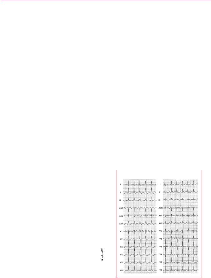
tooth waves in inferior leads and positive waves in V1 (Figure 10). In |
||||
. |
|||||
|
|
|
|
|
|
|
|
A |
|
B |
|
©ESC 2019
Figure 10 Counter-clockwise (A) and clockwise (B) atrial flutter with 2:1 atrioventricular conduction.
2019 September 13 on guest by 1093/eurheartj/ehz467/5556821.abstract/doi/10-article-com/eurheartj/advance.oup.https://academic from Downloaded

ESC Guidelines |
25 |
|
|
clockwise flutter, ECG flutter waves in inferior leads look positive and broad, and are frequently bimodal negative in V1.220,221 Typical atrial flutter has a strong reproducible anatomical dependence,222 resulting in the morphological reproducibility of the ECG. However, this well-recognized ECG pattern may be significantly changed when atrial activation has been modified, as it is in cardiac surgery involving atrial tissue, after extensive radiofrequency ablation, or in advanced atrial disease.223,224 Antiarrhythmic drugs may also modify the typical
ECG pattern.225 In these situations, an atypical ECG does not rule out a circuit of typical flutter using the CTI.226
Typical flutter is related to AF in clinical practice, with both being associated with similar clinical settings and coexisting in the same patients: AF may trigger atrial flutter, and after typical flutter ablation AF is frequent.23,227 229 Typical flutter may also frequently occur in patients treated for AF with class IC drugs or amiodarone. In this case, flutter rate may be reduced to <200 b.p.m. facilitating 1:1 AV conduction. The action of antiarrhythmic drugs on ventricular activation may result in wide QRS tachycardia.230 232
.
.
.
.
.
.
.
.
.
.
.
.
.
.
.
.
.
.
.
.
.
.
.
.
.
.
.
.
.
.
.
.
.
.
.
.
.
.
.
.
.
.
.
.
.
.
.
.
.
.
.
.
.
.
.
Beyond symptoms associated with high-rate and loss of atrial kick, reversible systolic dysfunction and subsequent TCM are not unusual.233,234
11.1.4.1.3 Acute therapy. Although the first step should be rate control when the ventricular rate is high, this may be difficult to achieve. AV nodal blocking drugs,235 238 including amiodarone applied mostly in HF or critically ill patients,239,240 may be of help, but cardioversion may be necessary (Figure 11).
In some cases, presenting with 2:1 AV block, the diagnosis of atrial flutter may not be obvious on the ECG. In these situations, i.v. adenosine may increase the degree of AV block and reveal the typical ECG pattern. However, adenosine can produce a rebound increase in AV conduction to 1:1 and may also precipitate AF.271,272 Thus, it should only be used if deemed necessary for diagnosis and resuscitation equipment is available.
Rate control should be the first step in very symptomatic patients with rapid ventricular rates. This is a particularly difficult goal in atrial
Recommendations for the therapy of macro2re-entrant atrial arrhythmias
|
Recommendations |
Classa |
Levelb |
|
|
|
|
|
|
|
Anticoagulation, as in AF, is recommended for patients with atrial flutter and concomitant AF.4 |
I |
B |
|
|
Patients with atrial flutter without AF should be considered for anticoagulation, but the threshold for initiation has not been |
IIa |
C |
|
|
established.241 247 |
|
||
|
|
|
|
|
|
Acute therapy |
|
|
|
|
Haemodynamically unstable patients |
|
|
|
|
Synchronized DC cardioversion is recommended for haemodynamically unstable patients.248,249 |
I |
B |
|
|
Haemodynamically stable patients |
|
|
|
|
i.v. ibutilide or i.v. or oral (in-hospital) dofetilide are recommended for conversion to sinus rhythm.250 257 |
I |
B |
|
|
Low-energy (<100 J biphasic) electrical cardioversion is recommended for conversion to sinus rhythm.248,249 |
I |
B |
|
|
High-rate atrial pacing is recommended for termination of atrial flutter in the presence of an implanted pacemaker or |
I |
B |
|
|
defibrillator.258 260 |
|
||
|
|
|
|
|
|
i.v. beta-blockers or non-dihydropyridine calcium channel blockers (verapamil or diltiazem) (i.v.), should be considered for control of |
IIa |
B |
|
|
rapid ventricular rate.235 238 |
|
||
|
|
|
|
|
|
Invasive and non-invasive high-rate atrial pacing may be considered for termination of atrial flutter.258,261 |
IIb |
B |
|
|
i.v. amiodarone may be tried if the above are not available or desirable.239,240 |
IIb |
C |
|
|
Propafenone and flecainide are not recommended for conversion to sinus rhythm.250 |
III |
B |
|
|
Chronic therapy |
|
|
|
|
Catheter ablation should be considered after the first episode of symptomatic typical atrial flutter.262,263 |
IIa |
B |
|
|
Catheter ablation is recommended for symptomatic, recurrent episodes of CTI-dependent flutter.262 264 |
I |
A |
|
|
Catheter ablation in experienced centres is recommended for symptomatic, recurrent episodes of non-CTI-dependent |
I |
B |
|
|
flutter.224,265 269 |
|
||
|
|
|
|
|
|
Catheter ablation is recommended in patients with persistent atrial flutter or in the presence of depressed LV systolic function due |
I |
B |
|
|
to TCM.233,234 |
|
||
|
|
|
|
|
|
Beta-blockers or non-dihydropyridine calcium channel blockers (verapamil or diltiazem, in the absence HFrEF) should be considered |
IIa |
C |
|
|
if ablation is not desirable or feasible.237,270 |
|
||
|
|
|
|
|
|
Amiodarone may be considered to maintain sinus rhythm if the above measures fail.263 |
IIb |
C |
|
|
AV nodal ablation with subsequent pacing (‘ablate and pace’), either biventricular or His-bundle pacing, should be considered if all the |
IIa |
C |
|
|
above fail and the patient has symptomatic persistent macro-re-entrant atrial arrhythmias with fast ventricular rates. |
|
||
|
|
|
|
|
|
|
|
|
|
i.v. verapamil and diltiazem are contraindicated in the presence of hypotension or HFrEF. i.v. beta-blockers are contraindicated in the presence of decompensated heart failure.
i.v. ibutilide, and i.v. and oral dofertilide are contraindicated in patients with prolonged QTc interval. i.v. amiodarone prolongs the QTc but torsades des pointes is rare.
AF = atrial fibrillation; AV = atrioventricular; CTI = cavotricuspid isthmus; DC = direct current; HFrEF = heart failure with reduced ejection fraction; i.v. = intravenous; LV = left ventricular; TCM = tachycardiomyopathy.
aClass of recommendation. bLevel of evidence.
2019 September 13 on guest by 1093/eurheartj/ehz467/5556821.abstract/doi/10-article-com/eurheartj/advance.oup.https://academic from Downloaded