Материал: Kaplan USMLE-1 (2013) - Anatomy
Section I • Histology and Cell Biology
Generalfeatures
•The colon is larger in diameter and shorter in length than is the small intestine. Fecal material moves from the cecum, through the colon
(ascending, transverse, descending, and sigmoidcolons), rectum, and anal canal.
•Three longitudinal bands of muscle, the teniae coli, constitute the outer layer. Because the colon is longer than these bands, pouching occurs, creating haustrabetween the teniae and giving the colon its characteristic "caterpillar" appearance.
•The mucosa has novilli,and mucus is secreted by short, inward-projecting colonic glands.
•Abundant lymphoid follicles are found in the cecum and appendix, and more sparsely elsewhere.
•The major functions of the colon are reabsorption offluid and electro lytes and temporary storage offeces.
Colonicmotility
•Peristalticwaves briefly open the normally closed ileocecal valve, passing a small amount of chyme into the cecum. Peristalsis also advances the chyme in the colon. Slow waves, approximately 2/min, are initiated at the ileocecal valve and increase to approximately 6/min at the sigmoid colon.
•Segmentation contractions mix the contents of the colon back and forth.
•Mass movement contractions are found only in the colon. Constriction of long lengths of colon propels large amounts of chyme distally toward the anus. Mass movements propel feces into the rectum. Distention of the rectum with feces initiates the defecation reflex.
Absorption
The mucosa of the colon has great absorptive capability. Na+ is actively trans ported withwaterfollowing, and K+ and HC03-are secreted into the colon.
Defecation
Feces: Contains undigested plant fibers, bacteria, inorganic matter, and water. Nondietary material (e.g., sloughed-off mucosa) constitutes a large portion of the feces. In normal feces, 30% of the solids may be bacteria. Bacteria synthesize vitamin K, B-complex vitamins, and folic acid; split urea to NH3; and produce small organic acids from unabsorbed fat and carbohydrate.
Defecation: Rectal distention with feces activates intrinsic and cord reflexes that cause relaxation of the internal anal sphincter (smooth muscle) and produce the urge to defecate. If the external anal sphincter (skeletal muscle innervated by the pudenda! nerve) is then voluntarily relaxed, and intra-abdominal pressure is in creased via the Valsalva maneuver, defecation occurs. Ifthe external sphincter is held contracted, the urge to defecate temporarily diminishes.
Gastrocolic reflex: Distention of the stomach by food increases the frequency ofmass movements and produces the urge to defecate. This reflex is mediated by parasympathetic nerves.
102 M EDICAL
Chapter 8 • Gastrointestinal System
Throughout the large intestine, the epithelium contains goblet, absorptive, and enteroendocrine cells. Unlike the small intestine, about half of the epithelial cells are mucus-secreting goblet cells, providing lubrication. The major function ofthe large intestine is fluid retrieval. Some digestion is still occurring, mainlythe break down ofcellulose by the permanent bacterial flora. Stem cells are in the lower part ofthe crypts.
Copyright McGraw-Hill Companies. Used withpermission.
Figure 1-8-9. Large intestine with crypts but no villi and many light-staining goblet cells interspersed among enterocytes
GASTROINTESTINAL GLANDS
Salivary Glands
The major salivary glands are all branched tubuloalveolar glands, with secretory acinithatdrain into ducts, which drain into the oral cavity. Acini contain serous, mucous, or both types ofsecretory cells, as well as myoepithelialcells, both sur rounded by a basal lamina. Serous cells secrete various proteins and enzymes. Mucous cells secrete predominantly glycosylated mucins. Plasma cells in the un derlying connective tissue, mainly in the parotid gland, secrete IgA molecules, which are transported across acinar and small ductal epithelial cells and secreted into saliva.
Clinical Correlate
Loss ofwater in the large intestine can be life-threatening in cholera and other intestinal infections. Irradiation and antimitotic compounds in cancer treatment destroy the rapidly dividing stem cells. Because the epithelium is not replaced, malabsorption and diarrhea develop until quiescent stem cells repopulate.
MEDICAL 103
Section I • Histology and Cell Biology
Table 1-8-4. Gastrointestinal Physiology
Appetite
Appetite is primarily regulated by 2 regions ofthe hypothalamus: a feeding center and a satiety center. Normally, the feeding center is active but is transiently inhibited bythe satiety center.
|
Hypothalamus |
|
||
|
Location |
Stimulation |
Destruction |
|
Feeding center |
Lateral |
|
Anorexia |
|
Feeding |
|
|||
|
hypothalamic |
|
|
|
|
area |
|
|
|
Satiety center |
Ventromedial |
Cessation of |
|
Hypothalamic |
|
nucleus of |
feeding |
|
obesity syndrome |
|
hypothalamus |
|
|
|
|
Hormones That May Affect Appetite |
|
||
Cholecystokinin |
• Released from I cells in the mucosa ofthe small intestine |
|||
(CCK) |
• CCK-A receptors are in the periphery |
|
||
|
|
|||
|
• CCK-B receptors are in the brain |
|
||
|
• Both reduce appetite when stimulated |
|
||
Calcitonin |
Released mainly from the thyroid gland |
|||
|
Has also been reported to decrease appetite by an |
|||
|
unknown mechanism |
|
||
Mechanical Distention
•Distention of the alimentary tract inhibits appetite, whereas the contractions of an empty stomach stimulate it.
•Some satiety is derived from mastication and swallowing alone.
Miscellaneous
Other factors that help to determine appetite and body weight include body levels of fat and genetic factors.
(Continued)
104 MEDICAL
Table 1-8-4. Gastrointestinal Physiology (Cont'd.) |
Chapter 8 • Gastrointestinal System |
||
|
|||
|
• |
Saliva |
|
Salivary glands |
Produce approximately 1.5 L/day of saliva |
|
|
Submandibular |
• |
Presence offood in the mouth; the taste, smell, sight, |
|
or thought of food; or the stimulation ofvagal afferents |
|
||
Parotid |
|
at the distal end ofthe esophagus increase production |
|
Sublingual |
|
|
|
|
of saliva |
|
|
|
|
|
|
Functions |
|
Initial triglyceride digestion (lingual lipase) |
|
|
|
Initial starch digestion (a-amylase) |
|
|
|
Lubrication |
|
Composition
Regulation
Ions: HC03-3x [plasma]; K+ 7 x[plasma];
Na+ 0.1 x [plasma]; o- 0.1 5 x [plasma]
Enzymes: a-amylase, lingual lipase
Hypotonic pH: 7-8
Flow rate: alters the composition
Antibacterial: lysozyme, lactoferrin, defensins, lgA
Parasympathetic |
i synthesis and secretion ofwatery |
|
|
saliva via muscarinic receptor |
-7 |
|
stimulation; (anticholinergics |
|
|
dry mouth) |
|
Sympathetic |
i synthesis and secretion ofviscous |
|
|
saliva via 13-adrenergic receptor |
|
|
stimulation |
|
The·ducts that drain the glands increase in size and are lined by an epithelium that transitions from cuboidal to columnar to pseudostratified to stratified co lumnar cells. The smallest ducts, intercalated ducts, have myoepithelial cells; the next larger ducts, striated ducts, have columnar cells with basal striations, caused by basal infolding of cell membranes between prominent mitochondria. These columnar cells make the saliva hypotonic by transporting Na and Cl ions out of saliva back into the blood.
|
105 |
MEDICAL |
Section I • Histology and Cell Biology
Clinical Correlate
The parotid gland is the major site of the mumps and rabies viruses that are transmitted in saliva.
Benign tumors most frequently appear at the parotid gland; their removal
is complicated by the facial nerve traversing the gland.
CopyrightMcGraw-Hi/I Companies. Used withpermission.
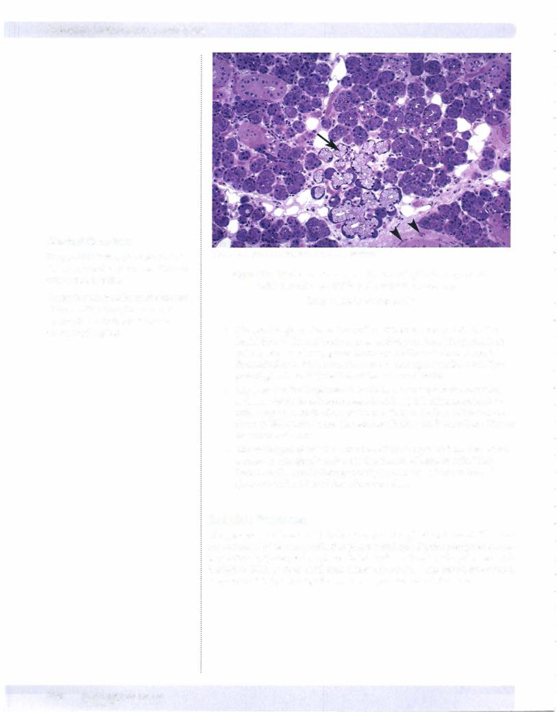
Figure 1-8-10. Submandibular with a mix of light-staining mucus acini (arrow) adjacent to dark-staining serous acini
Small vessels (arrowheads)
•The parotid glands lie on the surface ofthe masseter muscles in the lateral face, in front of each external auditory meatus. The parotids are entirely serous salivary glands that drain inside each cheek through Stensen's ducts which open above the second upper molar tooth. The parotid glands contribute 25% of the volume of saliva.
•The submandibular glands lie inside the lower edge of the mandible, and are mixed serous/mucous glands with a predominance ofserous cells. They drain in the floor of the mouth near the base of the tongue through Wharton's ducts. The submandibular glands contribute 70% of the volume of saliva.
•The sublingual glands lie at the base of the tongue, and are also mixed serous/mucous glands with a predominance ofmucous cells. They drain into the mouth through multiple small ducts, The sublingual glands contribute 5% of the volume of saliva.
Exocrine Pancreas
The pancreas is a branched tubuloacinar exocrine gland with acini. The acini are composed ofsecretory cells that produce multiple digestive enzymes includ ing proteases, lipases, and amylases. Acinar cells are functionally polarized, with basophilic RER at their basal ends below the nucleus, and membrane-bound, enzyme-containing eosinophilic zymogen granules toward their apex.
1 06 MEDICAL