Материал: Kaplan USMLE-1 (2013) - Anatomy
Chapter 8 • Gastrointestinal System
Copyright McGraw-Hi/I Companies. Usedwithpermission.
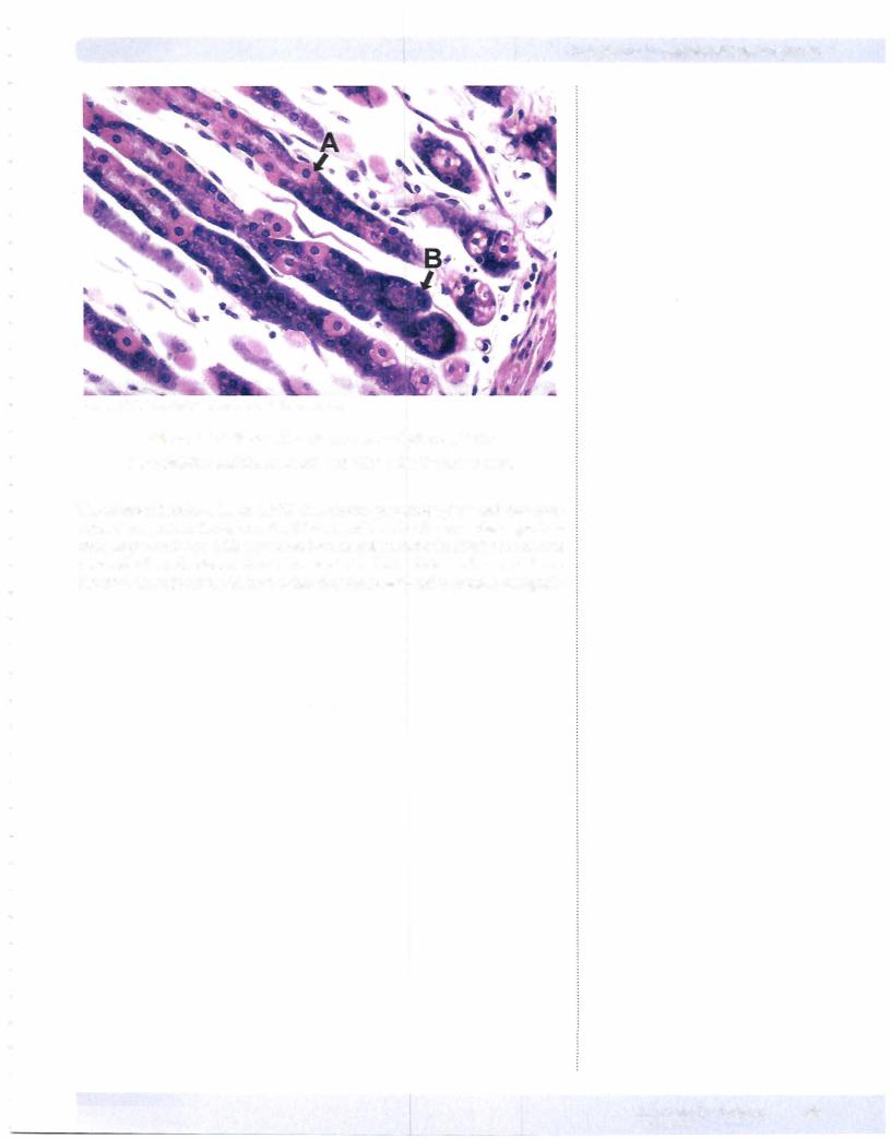
Figure 1-8-4. Stomach near the base of gastric glands
Pale-staining parietal cells (A) and chief cells (B) are shown.
The enteroendocrine cells or APUD cells (amine precursor uptake and decarbox ylation) are present throughout the GI tract and are also found in the respiratory tract. They constitute a diffuse neuroendocrine system that collectively accounts for more cells than allother endocrine organs in the body. Enteroendocrine cells are dispersed throughout the GI tract so thattheycan receive and transmit local signals.
MEDICAL 93
Section I • Histology and Cell Biology
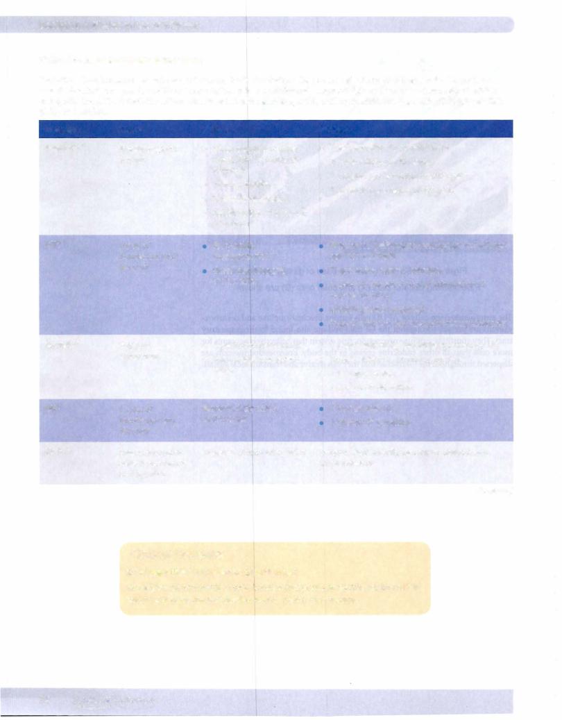
Table 1-8-3. Gastrointestinal Hormones
Gastrointestinal hormones are released into the systemic circulation after physiologic stimulation (e.g., by food in gut), can exert their effects independent ofthe nervous system when administered exogenously, and have been chemically identified and synthesized. The 5 gastrointestinal hormones include secretin, gastrin, cholecystokinin (CCK), gastric inhibitory peptide (GIP), and motilin.
Hormone |
Source |
Stimulus |
|
Gastrin*• * |
G cells of gastric |
• |
Small peptides, amino |
|
antrum |
• |
acids, Ca2+ in lumen of |
|
|
stomach |
|
|
|
Vagus (via GRP) |
|
|
|
• |
Stomach distension |
• Inhibited by: W in lumen of antrum
Actions
•i HCl secretion by parietal cells
•Trophic effects on GI mucosa
•i pepsinogen secretion by chief cells
•i histamine secretion by ECL cells
CCK* |
I cells of |
Fatty acids, |
|
duodenum and |
monoglycerides |
|
jejunum |
Small peptides and |
|
|
|
|
|
amino acids |
Secretint |
S cells of |
• J, pH in duodenal lumen |
|
duodenum |
• Fatty acids in duodenal |
|
|
|
|
|
lumen |
GIPt |
K cells of |
Glucose, fatty acids, |
|
duodenum and |
amino acids |
|
jejunum |
|
Motilin |
Enterochromaffin |
Absence of food for >2 hours |
|
cells in duodenum |
|
|
and jejunum |
|
Stimulates gallbladder contraction and relaxes sphincter of Oddi
i pancreatic enzyme secretion
Augments secretin-induced stimulation of pancreatic HC03-
Inhibits gastric emptying
Trophic effect on exocrine pancreas/gallbladder
•i pancreatic HC03- secretion (neutralizes W)
•Trophic effect on exocrine pancreas
•i bile production
•J, gastric W secretion
i insulin release
J, gastric W secretion
Initiates MMC motility pattern in stomach and small intestine
(Continued)
Clinical Correlate
*Zollinger-Ellison syndrome (gastrinoma)
Non-p islet-cell pancreatic or pyloric tumor that produces gastrin, leading to i in gastric acid secretion and development of peptic ulcer disease
94 MEDICAL
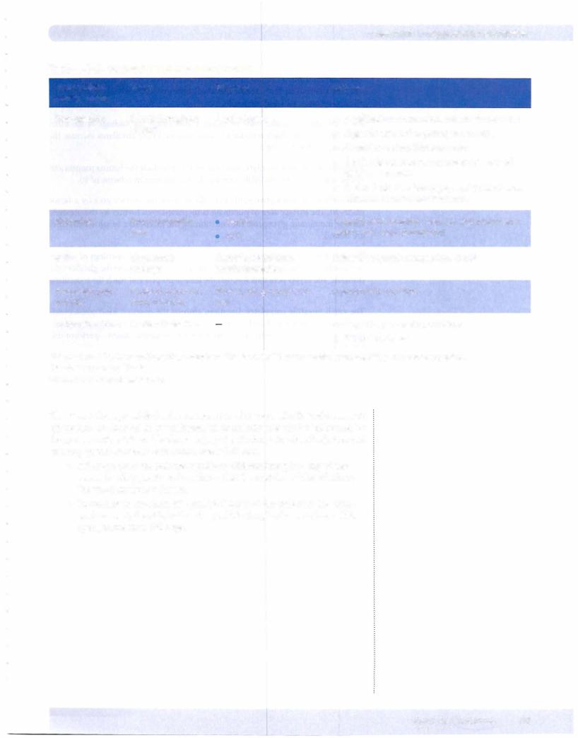
Table 1-8-3. Gastrointestinal Hormones (Cont'd.)
PARACRINES/ |
Source |
Stimulus |
NEUROCRINES |
|
|
Somatostatin |
D cells throughout |
|
GI tract |
Histamine |
Enterochromaffin |
|
cells |
GRP |
Vagal nerve |
|
endings |
Pancreatic poly- |
F cells of pancreas, |
peptide |
small intestine |
Enteroglucagon |
L cells of intestine |
J, pH in lumen
Gastrin
ACh
Cephalic stimulation, gastric distension
Protein, fat, glucose in lu- men
Chapter 8 • Gastrointestinal System
Actions
•j, gallbladder contraction, pancreatic secretion
•J, gastric acid and pepsinogen secretion
•J, small intestinal fluid secretion
•j, ACh release from the myenteric plexus and decreases motility
•J, a-cell release of glucagon, and P-cell release of insulin in pancreatic islet cells
t gastric acid secretion (directly, and potentiates gastrin and vagal stimulation)
Stimulates gastrin release from G cells
J, pancreatic secretion
•J, gastric, pancreatic secretions
•t insulin release
Abbreviations: CCK, cholecystokinin; ECL, enterochromaffin-like cells; GIP, gastric inhibitory peptide; GRP, gastrin-releasing peptide. *Member of gastrin-CCK family
tMember of secretin-glucagon family
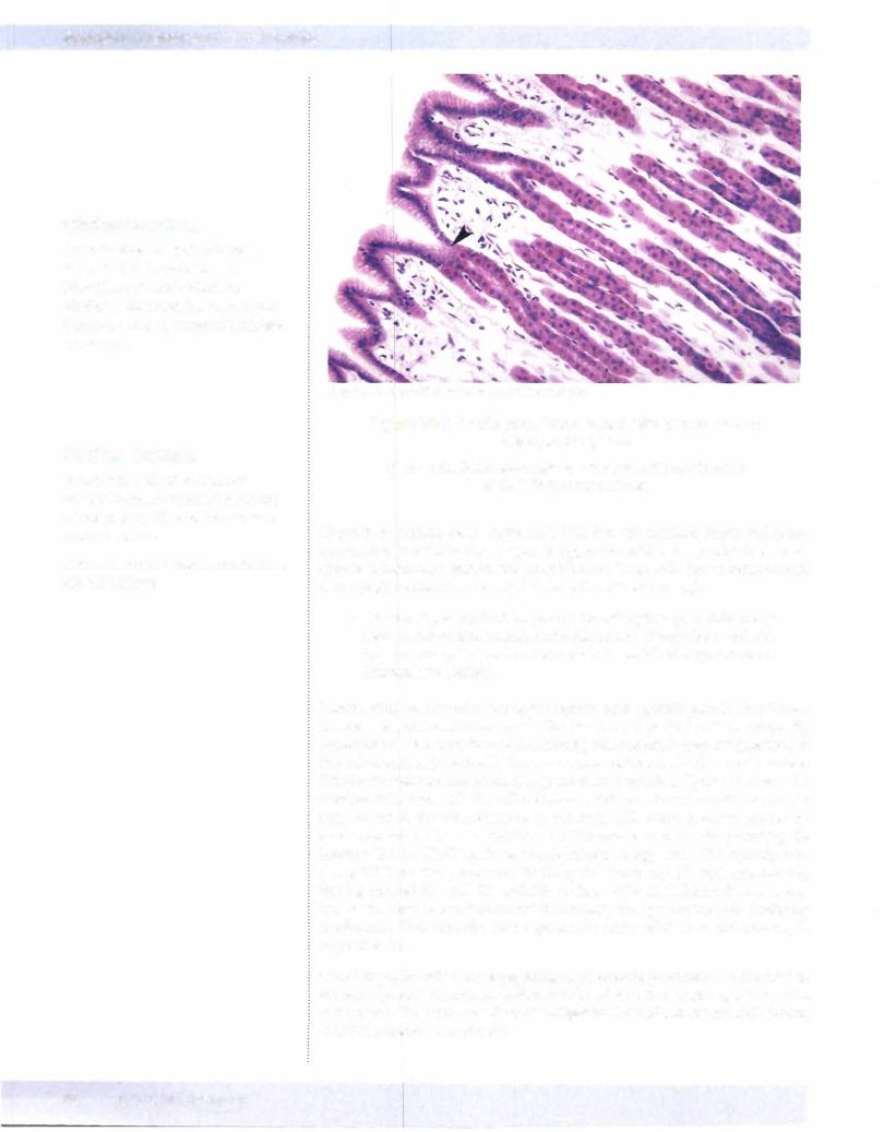
The stemcells responsible for the regeneration of all types of cells in the stomach epithelium are located in the isthmus. Their mitotic rate can be influenced by the presence of gastrin and by damage (aspirin, alcohol, bile salt reflux). Renewal of many gastric epithelial cells occurs every 4-7 days.
•Although the stem cells are capable of differentiating into any of the stomach cell types, there is evidence that the position of the cell along the gland influences its fate.
•In contrast to the short life span (4-5 days) of the cells near the acidic environment, the chief cells-deep within the glands-may have a life span greater than 1 90 days.
MEDICAL 95
Section I • Histology and Cell Biology
Small Intestine
The small intestine is tubular in shape and has a total length of about 21 feet. The effective internal surface area of the small intestine is greatly increased by the plicae circulares, villi and microvilli.
Plicae circulares (circular folds or valves of Kerckring) are foldings of the inner surface that involve both mucosa and sub-mucosa. Plicae circulares increase the surface area by a factor of 3.
Villi arise above the muscularis mucosae and they include the lamina propria and epithelium of the mucosa. Villi increase the surface area by a factor of 10.
Microvilli of the absorptive epithelial cells increase the surface area by a factor of20-30. 'Ihe surface area of microvilli is increased even further by the presence surface membrane glycoproteins, constituting the glycocalyx to which enzymes are bound.
The luminal surface ofthe small intestine is perforated by the openings of numer ous tubular invaginations (the crypts of Lieberkiihn) analogous to the glands of the stomach. The crypts penetrate through the lamina propria and reach the muscularis mucosae.
The small intestine completes digestion, absorbs the digested food constituents (amino acids, monosaccharides, fatty acids) and transports it into blood and lym phatic vessels. Three subdivisions-duodenum, jejunum, ileum-perform dif ferent digestive functions.
For carbohydrates, pancreatic amylase completes the hydrolysis of poly saccharides to disaccharides that is initiated by salivary amylase in the mouth.
For proteins, pancreatic enzymes, trypsin, chymotrypsin, elastase and carboxypeptidase mediate further digestion to small peptide fragments.
•For dietaryfat, pancreatic lipase hydrolyzes fat to free fatty acids and monoglycerides. These combine with bile salts to form micelles. The micelles diffuse across the surface membrane of microvilli and then to the smooth endoplasmic reticulw11 (SER) where triglycerides are resynthesized and processed into chylomicrons in the Golgi apparatus.
Chylomicrons are transported out the basal surface into the lamina pro pria and diffuse into the lacteals in the center of the villus.
Sugars and amino acids are absorbed through the enterocytes and then transported to the capillary network immediately below the epithelium.
The small intestine participates in the digestion and absorption of nutrients. It has specialized villi on the epithelial surface to aid in this function.
The duodenum is the proximal pyloric end of the small intestine. Distal to the duodenum is the jejunum, and then the ileum.
In the small intestine, the chyme from the stomach is mixed with mucosa! cell secretions, exocrine pancreatic juice, and bile.
96 M EDICAL