Материал: Kaplan USMLE-1 (2013) - Anatomy
Chapter 8 • Gastrointestinal System
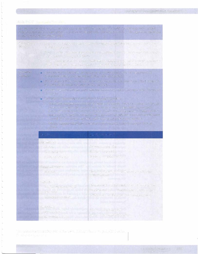
Table 1-8-5. Pancreatic Secretions
The exocrine secretions ofthe pancreas are produced by the acinar cells, which contain numerous enzyme containing granules in their cytoplasm, and by the ductal cells, which secrete HC03-. The secretions reach the duodenum via the pancreatic duct.
Bicarbonate |
• HC03in the duodenum neutralizes HCl in chyme entering from the stomach. This also |
|||||
(HC03-) |
||||||
deactivates pepsin. |
|
|
|
|
||
|
• When W enters the duodenum, S cells secrete secretin, which acts on pancreatic ductal |
|||||
|
cells to increase HC03production. |
|
|
|||
|
• HC03is produced by the action of carbonic anhydrase on C02 and H20 in the pancreatic |
|||||
|
ductal cells. HC03is secreted into the lumen ofthe duct in exchange for o-. |
|||||
Pancreatic |
Approximately 1 5 enzymes are produced by the pancreas, which are responsible for |
|||||
enzymes |
digesting proteins, carbohydrates, lipids, and nucleic acids. |
|||||
|
When small peptides, amino adds, and fatty acids enter the duodenum, CCK is released |
|||||
|
by I cells, stimulating pancreatic enzyme secretion. |
|
|
|||
|
ACh (via vagovagal reflexes) also stimulates enzyme secretion and potentiates the action |
|||||
|
of secretin. |
|
|
|
|
|
|
Protection ofpancreatic acinar cells against self-digestion: |
|||||
|
- Proteolytic enzymes are secreted as inactive precursors, which are activated in the gut |
|||||
|
lumen. For example, the duodenal brush border enzyme, enterokinase, converts |
|||||
|
trypsinogen to the active enzyme, trypsin. Trypsin then catalyzes the formation of more |
|||||
|
trypsin and activates chymotrypsinogen, procarboxypeptidase, and prophospholipases |
|||||
|
A and B. Ribonucleases, amylase, and lipase do not exist as proenzymes. |
|||||
|
- Produce enzyme inhibitors to inactivate trace amounts of active enzyme formed |
|||||
|
within. |
|
|
|
|
|
|
Enzyme |
|
Reaction Catalyzed |
|
|
|
|
Proteases: |
|
Proteins peptides |
|
||
|
|
|
||||
|
Trypsin |
|
|
|||
|
Chymotrypsin |
|
Proteins peptides |
|
||
|
Carboxypeptidase |
|
Peptides amino acids |
|||
|
Polysaccharidase: |
|
|
|
|
|
|
Amylase |
|
Starch and glycogen |
maltose, maltotriose, and |
||
|
|
a-limit dextrins |
|
|||
|
Lipases: |
|
Phospholipids phosphate,fatty acids, and glycerol |
|||
|
Phospholipases A and B |
|
||||
|
Esterases |
|
Cholesterol esters free cholesterol and fatty acids |
|||
|
Triacylglycerol lipases |
|
Triglycerides fatty acids and monoglycerides |
|||
|
Nucleases: |
|
RNA ribonucleotides |
|||
|
Ribonuclease |
|
||||
|
Deoxyribonuclease |
|
DNA deoxyribonucleotides |
|||
The endocrine-producing cells of the islets ofLangerhans are embedded within the exocrine pancreas.
MEDICAL 107
Section I • Histology and Cell Biology
Copyright McGraw-Hill Companies. Used with permission.
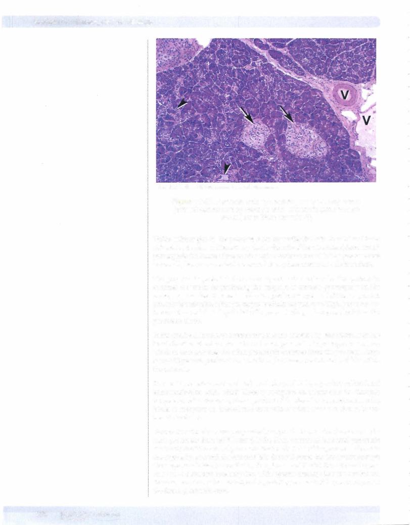
Figure 1-8-11. Pancreas with light-staining islets of Langerhans (arrows) surrounded by exocrine acini with ducts (arrowheads) and adjacent blood vessels (V)
Unlike salivary glands, the pancreas lacks myoepithelial cells in acini and lacks striated ducts. Also, unlike salivary glands, the cells ofthe intercalated. ducts extend partially into the lumen ofpancreatic acini as centroacinarcells. The pancreas does not usually have mucinous cells in acini, but may have mucinous cells in its ducts.
The pancreas is protected from auto-digestive destruction by the proteolytic enzymes it secretes by producing the enzymes as inactive proenzymes in the zymogen granules. Pancreatis cells also produce trypsin inhibitor to prevent proteolytic activation ofthe proenzymes within the pancreas. Tight junctions be tween acinar and ductal epithelial cells prevent leakage of enzymes back into the pancreatic tissue.
In the duodenal lumen, pancreatic enzymes are activated by enterokinase in the brush border of enterocytes. This activates pancreatic trypsinogen to trypsin, which in turn activates the other proteolytic enzymes from the pancreas. Amy lase and lipase are produced in active form, but have no substrate available within the pancreas.
Pancreatic secretion is stimulated by cholecystokinin, a product of duodenal enteroendocrine cells, which binds to receptors on acinar cells to stimulate enzyme secretio. Secretin, also a product of duodenal enteroendocrine cells, binds to receptors on intercalated duct cells to stimulate secretion of bicar bonate and water.
Pancreatic acini drain via progressively larger ducts into the duodenum. The main pancreatic duct (ofWirsung) is the distal portion of the dorsal pancreatic duct that joined the ventral pancreatic duct in the head ofthe pancreas. The main duct typicallyjoins with the common bile duct and enters the duodenum through the ampulla ofVater (controlled by the sphincter ofOddi). Sometimes the pan creas has a persistent accessory duct with separate drainage into the duodenum, the accessory duct ofSantorini, and a persisting remnant ofthe proximal part of the dorsal pancreatic duct.
108 M EDICAL
Section I • Histology and Cell Biology
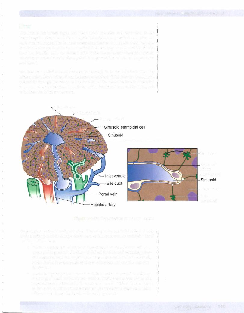
The hepatic artery and portal vein enter and the common hepatic duct exits the liverin the hepatic hilum. Within the liver, branches ofthe hepatic artery, portal vein, and bile duct tend to run together in thin connective tissue bands. When seen in cross section, these 3 structures and their connective tissue are called a portal triad or portal tract. Blood from portal vein and hepatic artery branches both flow through and mix in hepatic sinusoids that run between cords or plates of hepatocytes. After passing by hepatocytes, the sinusoidal blood flows into he patic venules, which form progressively larger branches draining into the right and left hepatic veins which drain into the inferior vena cava.
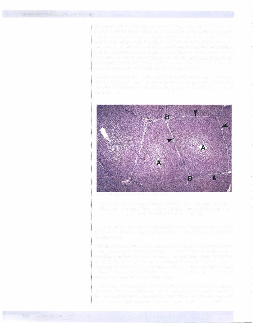
A classic hepatic lobule is a hexagonal structure with a portal tract at each corner ofthe hexagon and a centralvein in the center ofthe hexagon. Blood flowis from the triads into the central vein and bile flow is opposite, from the central vein to the triads.
CopyrightMcGraw-Hill Companies. Usedwith permission.
Figure 1-8-13. Liver lobules with central veins (A) in the center of each lobule and connective tissue (arrowheads) separating each lobule and portal triads at each point of the lobule (B)
A portal lobule is a triangular structure with a central vein at each corner and a portal tract in the center. Bile flows from the periphery ofthe portal lobule into the central triad.
A hepatic acinus is based on blood flow from the hepatic arterybranches to cen tral veins. As hepatic arterial blood flow enters the sinusoids from side branches extending away from the center of the hepatic triad (rather than directly from the triad), the center of the acinus is conceived of as centered on such a branch extending out from a triad (or between 2 triads) and ending at 2 nearby central veins, resulting in a roughly elliptical structure with portal tracts at the 2 furthest poles and 2 central veins at the 2 closest edges.
In the acinus, the hepatocytes receiving the first blood flow (and the most oxy gen and nutrients) are designated zone l, while those receiving the last blood flow (and least oxygen and nutrients) are near the central veins and designated zone 3. Zone 2 hepatocytes are in between zones 1 and 3. This model helps to
110 MEDICAL
Chapter 8 • Gastrointestinal System
explain the differential effect on hepatocytes of changes in blood flow, oxy genation, etc. Zone 3 is most susceptible to injury by decreased oxygenation of blood or decreased blood flow into the liver (as well as stagnation of blood drainage out ofthe liver due to congestive heart failure).
The metabolic activity of hepatocytes varies within the zones of the acinus. Zone 1 hepatocytes are most involved in glycogen synthesis and plasma pro tein synthesis (albumin, coagulation factors and complement components). Zone 3 cells are most concerned with lipid, drug, and alcohol metabolism and detoxification.
Different functions of the hepatocyte are concentrated in different organelles.
•Rough endoplasmic reticulum (RER) is responsible for protein synthesis
(e.g., albumin, coagulation. factors, complement components, and lipo proteins).
•Smooth endoplasmic reticulum (SER) has many enzymes associated with its membranes and is responsible for synthesis of cholesterol and bile acids, conjugation (solubilization) of bilirubin and lipid soluble drugs, formation of glycogen under control of insulin (glycogen rosettes are often associated with SER) and breakdown of glycogen to glucose (glycogenolysis) under the control of glucagon and epinephrine, and detoxification of lipid soluble drugs (e.g., phenobarbital), including via the microsomal enzyme oxidizing system (MEOS).
•The Golgi apparatus is responsible for glycosylation of proteins and packaging some proteins for secretion.
•Lysosomes are responsible for degradation of aged plasma glycoproteins taken up from the blood.
•Peroxisomes are responsible for breakdown of hydrogen peroxide.
Ito cells (stellate cells) are mesenchymal cells that live in the space of Disse. They contain fat and are involved in storage of fat-soluble vitamins, mainly vitamin A.
Bile formation by hepatocytes serves both an exocrine and excretory function. Bile salts secreted into the duodenum aid in fat emulsification and absorption, as well as excretion of endogenous metabolites (bilirubin) and drug metabolites that cannot be excreted by the kidney. Bile consists ofa mixture ofbilesalts (con jugated bile acids), conjugated bilirubin (and other conjugated endogenous or drug metabolites), cholesterol, phospholipids, electrolytes, and water.
Bile acids are synthesized from cholesterol by hepatocytes, and subsequently conjugated in SER to produce bile salts. Specific transporters at the bile cana liculus secrete bile salts into bile Within the gut lumen some bile salts are par tially metabolized by gut bacteria to produce other bile salts (deoxycholic and lithocholic acid). All ofthese bile salts can be reabsorbed, principally in the small intestine, and recycled in bile (the enterohepatic circulation ofbile).
The liver excretes bilirubin, a metabolic breakdown product of hemoglobin from old red blood cells. Bilirubin is not very water-soluble, and is transported in plasma bound to protein, chiefly albumin. Hepatocytes have specific transport proteins that import bilirubin into their cytoplasm, where the hepatocytes then "solubilize" the bilirubin by conjugating it, chiefly to glucuronic acid. This solu bilized form ofbilirubin is then secreted into bile by a specific transport system at the canaliculus.
Clinical Correlate
When stimulated during liver injury, Ito cells may release type I collagen and other matrix components into the
space of Disse, contributing to scarring ofthe liver in some diseases (cirrhosis due to ethanol). This may lead to the development of portal hypertension, portacaval anastomoses, and esophageal or rectal bleeding.
Clinical Correlate
Disturbance of the balance in the components of bile can lead to precipitation of one or more ofthe bile components, resulting in stone (or calculus) formation or lithiasis in the gallbladder and/or bile ducts.
MEDICAL 111