Материал: Kaplan USMLE-1 (2013) - Anatomy

Chapter 9 • Urinary System
Organization
The cortex is divided into lobules, and contains nephron elements mixed with vascular elements and stroma (a small amount ofconnective tissue). Atthe center ofeach lobule is amedullaryray, containingtubulesthatare parallelto each other and oriented radially in the cortex. The tubules in themedullaryrays are continu ous with those in the medulla. Along the 2 edges of each lobule are glomeruli, located along one or 2 rows. Radially oriented arterioles and venules with a large lumen are located at the edges ofthe lobules.
The medulla is comprised of radially arranged straight tubules which run from cortex to papilla, vascular elements, and stroma (a small amount of connective tissue). The medulla is divided into 2 zones. A wide strip in proximity to the cor tex, the outermedullacontains profiles oftubuleswith different appearances. The inner medullahas fewer profiles ofsimilar tubes.
Blood Circulation
The renal artery enters the kidney at the hilum, near the ureter. The artery branches into interlobar arteries, which travel to the medulla-cortex border re maining outside the medullarypyramids, The vessels branch into arcuate arteries (and veins) that follow the edge of the cortex. The arcuate arteries branch into interlobular arterioles that travel tangentially in the cortex at the edges of the lobules. Intralobular arterioles, feeding the glomeruli, branch offthe interlobular arterioles at each renal corpuscle.
The kidneys receive 25% oftotal cardiac output, 1,700 liters in 24 hours. Each in tralobular arteriole enters a renal corpuscle atthe vascular pole as afferent arteri ole and forms a convolutedtuft ofcapillaries (the glomerulus). A second arteriole (the efferent arteriole) exits the corpuscle. This is a unique situation due to the fact that the pressure remains high in the glomerulus in order to allow filtration.
The efferent arterioles carrying blood out ofthe glomeruli make a second capil larybed. This second capillarybed has lowerblood pressure than the glomerulus and it connects to venules at its distal end. The arteriole-capillary-arteriole-cap illary-vein sequence in the kidney is unique in the body. The efferent arterioles from glomeruli in the upper cortex divide into a complex capillary system in the cortex.
NEPHRON
The functional unit within the kidney is the nephron. Each kidney contains 1 - 1 .3 million nephrons. Nephrons connect to collecting ducts, and collecting ducts receive urine from several nephrons and converge with each other before opening to and letting the urine flow out of the kidney. The nephron and the collecting duct form the uriniferous tubule.
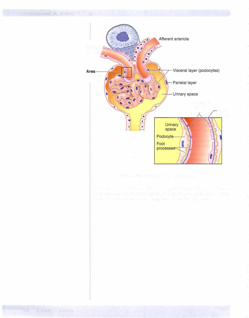
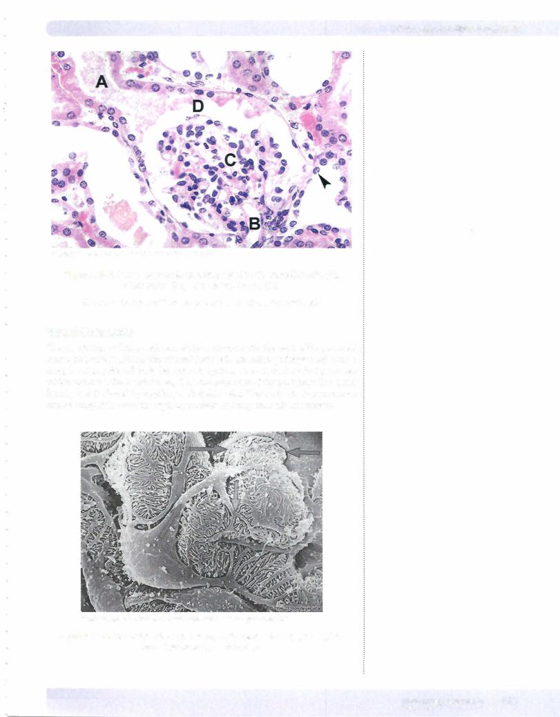
The nephron is a tube about 55 mm in length in the human kidney. It starts at one end with Bowman's capsule, which is the enlarged end ofthe nephron. Bowman's capsule has been invaginated by a tuft of capillaries of the glomerulus so that it has 2 layers: the visceral layer is in direct contact with the capillary endothelium, and the parietal layer surrounds an approximately spherical urinary space. Bow man's capsule and glomerulus ofcapillaries form a renal corpuscle.
MEDICAL 117
Section I • Histology and Cell Biology
Urinary (Bowman's) space
Podocyte foot processes
Podocyte
Capillary endothelial
RBC
From the IMC, @ 2010 DxR Development Group, Inc. All rights reserved.
Clinical Correlate
The absence of nephrin protein renders podocytes incapable of forming foot processes and slit diaphragms, and results in a congenital nephrotic syndrome, NPHSl.
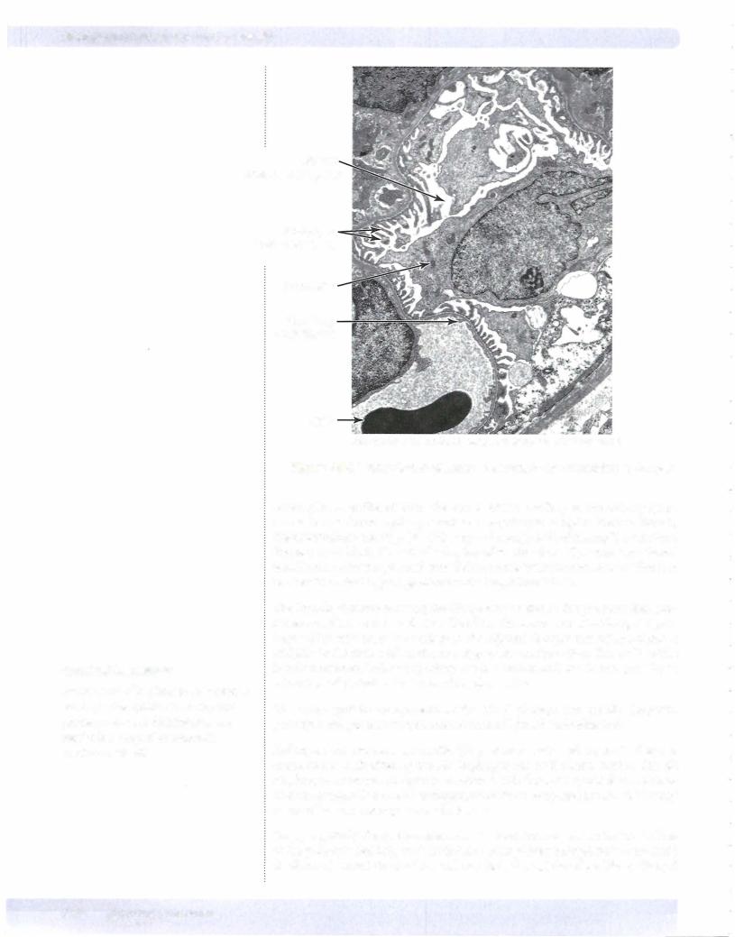
Figure 1-9-6 Transmission electron micrograph demonstrating podocytes
Blood plasma is filtered from the lumen of the capillary to the urinary space across the combined capillary endothelium-podocyte complex. Fenestrations in the endothelium are large (50-100 nm) and occupy 20% of the capillary surface. Fenestrations block the exit of cells, but allow free flow of plasma. The shared basal lamina ofpodocytes and endothelium constitutes the first, coarser filtration barrier; it blocks the passage of molecules larger than 70 kD.
The thin diaphragms covering the slit openings between the podocyte foot pro cesses constitutes a more selective filter. The slits are composed ofelongated pro teins which arise from the surface of the adjacent foot process cell membranes and join in the center of the slit, in a zipper-like configuration. The width of the junction between 2 adjacent podocytes varies between 20 and 50 nm, possibly as a function ofperfusion pressures ofthe glomerulus.
The major protein components of the slit diaphragm are specific (nephrin, podocyn) and generic components of other cell junctions (cadherins).
Podocyte foot processes are motile (they contain actin and myosin). They are connected to each other by the slit diaphragm and to the basal lamina. The slit diaphragm molecular complex is associated with the actin cytoskeleton. Altera tions in composition and/or arrangement of these complexes are found in many forms ofhuman and experimental diseases.
Large, negatively charged complexes in the basal lamina and the lateral surfaces ofthe podocyte feet help to slow diffusion ofnegatively charged molecules (such as albumin) across the lamina, and may help the uptake of positively charged
120 MEDICAL

Chapter 9 • Urinary System
molecules by binding them. The viscous gel consistency ofthe basal lamina is also a factor in retarding the diffusion ofmacromolecules.
Proximal Convoluted Tubule
The proximal convoluted tubule (PCT) opens at the urinary pole of Bowman's capsule. The PCT follows a circuitous path and ends with a straight segment that connects to the loop of Henle. PCT cells are tall, and they have a pink cytoplasm, long apical microvilli, and extensive basal invaginations. Numerous large mi tochondria are located between the basal invaginations. The lateral borders of adjacent cells are extensively interdigitated. These characteristics are typical of cells involved in active transport. The lumen ofthe PCT is frequently clouded by microvilli which do not preserve well during the histologic preparation process.
Loop of Henle
The loop of Henle has a smaller diameter than the PCT and has descending and ascending limbs which go in opposite directions. Some loops of Henle have a wider segment before the distal tubule. The straight and convoluted segments of the distal convoluted tubule (DCT) follow. The straight portions of the PCT and DCT have traditionally been assigned to the loop ofHenle (constituting the thick ascending and descending limbs) but they are now thought to be part ofthe PCT and DCT to which they are more similar. The special disposition of the loops of Henle descending and ascending branches, coupled with their specific transport and permeability properties, allow them to operate as "countercurrent multipli ers;' creating a gradient ofextracellular fluid tonicity in the medulla. This is used to modulate urine tonicity and finalvolume.
Distal Convoluted Tubule
The DCT comes back to make contact with its own glomerulus, and then con nects to the collecting tubule, which receives urine from several nephrons and is open at its far end. The epithelium of DCT, loops of Henle, and collecting ducts have variable thicknesses and more or less well-defined cell borders. Some have limited surface microvilli. In general, these tubes either do much less active trans port than the PCT or are involved only in passive water movements.
Collecting Ducts
Collecting ducts are linedbyprincipal cells and intercalated cells. The cell outline of these cells is more distinct than that of the PCT or the DCT. Principal cells respond to aldosterone.
Mesangial Cells
Mesangial cells (also known as Polkissen or Lacis cells) are located between capil laries, under the basal lamina but outside the capillary lumen. There is no basal lamina between mesangial and endothelial cells. Mesangial cells are phagocytic and may be involved in the maintenance of the basal lamina. Abnormalities of mesangial cells are detected in several diseases resulting in clogged and/or dis torted glomeruli.
Note
Renal cortex and medullary fibroblasts (interstitial cells) produce erythropoetin.
Note
Diuretics act by inhibiting Na+ resorption, leading to an increase in Na+ and water excretion.
MEDICAL 121