Chapter 10 • Male Reproductive System
Sertoli Cells and the Blood-Testis Barrier
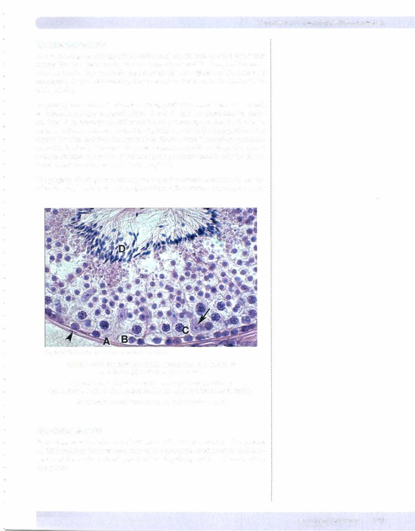
Sertoli cells are tall columnar epithelial cells. These multifunctional cells are the predominant cells in the seminiferous tubule prior to puberty and in elderly men but comprise only 10% of the cells during times of maximal spermatogenesis.
•Irregular in shape; the base adheres to the basal lamina and the apical end extends to the lumen. The nucleus tends to be oval with the long axis oriented perpendicular to the basement membrane.
•The cytoplasmic extensions make contact with neighboring Sertoli cells via tight junctions, forming the blood-testis barrier by separating the seminiferous tubule into a basal and an adlumenal compartment.
•Have abundant smooth endoplasmic reticulum, some rough endoplas mic reticulum, a well-developed Golgi complex, lysosomes, microtu bules, and microfilaments. Microtubules and microfilaments likely func tion to aid in the release of the spermatozoa.
Do not divide during the reproductive period.
Support, protect, and provide nutrition to the developing spermatozoa. During spermiogenesis, the excess spermatid cytoplasm is shed as resid ual bodies that are phagocytized by Sertoli cells. They also phagocytize germ cells that fail to mature.
•Secrete androgen-binding protein that binds testosterone and dihy drotestosterone. High concentrations of these hormones are essential for normal germ-cell maturation. The production of androgen-binding protein is stimulated by follicle-stimulatinghormone (FSH receptors are on Sertoli cells).
Secrete inhibin, which suppresses FSH synthesis.
Produce anti-Mullerian hormone during fetal life that suppresses the development of female internal reproductive structures.
The blood-testis barrier is a network of Sertoli cells which divides the seminif erous tubule into a basal compartment (containing the spermatogonia and the earliest primary spermatocytes) and an adlumenal compartment (containing the remaining spermatocytes and spermatids). The basal compartment has free ac cess to material found in blood, while the more advanced stages of spermato genesis are protected from blood-borne products by the barrier formed by the tight junctions between the Sertoli cells. The primary spermatocytes traverse this barrier by a mechanism not yet understood.
Interstitial Tissues ofthe Testis
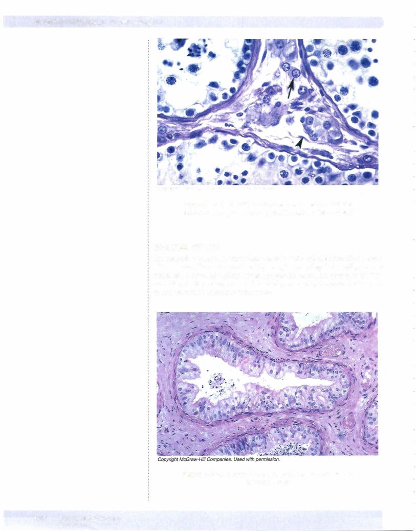
The interstitial tissue lying between the seminiferous tubules is a loose network of connective tissue composed of fibroblasts, collagen, blood and lymphatic ves sels, and Leydig cells (also called interstitial cells). The Leydig cells synthesize testosterone.