Материал: Kaplan USMLE-1 (2013) - Anatomy
Cell Biology and Epithelia |
1 |
NUCLEUS
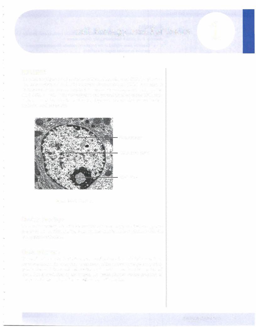
The nucleus (Figure 1-1-1) is the site of deoxyribonucleic acid (DNA) replication and transcription of DNA into precursor ribonucleic acid (RNA) molecules. It contains allof the enzymes required for replication and repair of newly synthe sized DNA, as well as for transcription and processing of precursor RNA mol ecules. It is enclosed by the nuclear envelope and contains the nuclear lamina, nucleolus, and chromatin.
Euchromatin
Heterochromatin
Nucleolus
Figure 1-1-1. Nucleus
Nuclear Envelope
The nuclear envelope is a double membrane containing pores that are approxi mately 90 nm in diameter. The outer nuclear membrane is continuous with the endoplasmic reticulum.
Nuclear Lamina
The nuclear lamina is a latticelike network of proteins that include lamins. Lam ins attach chromatin to the inner membrane of the nuclear envelope and partici pate in the breakdown and reformation of the nuclear envelope during the cell cycle.
mitosis initiates nuclear disassembly into smallvesicles.
MEDICAL 3

Section I • Histology and Cell Biology
Nucleolus
The nucleolus is responsible for ribosomal RNA (rRNA) synthesis and ribosome assembly. It contains 3 morphologically distinct zones:
•Granular zone-found at the periphery; contains ribosomal precursor particles in various stages of assembly.
•Fibrillar zone-centrally located; contains ribonuclear protein fibrils.
•Fibrillar center-containsDNA that is not being transcribed.
Chromatin
Chromatin is a complex of DNA, histone proteins, and nonhistone proteins.
•DNA-a double-stranded helical molecule that carries the genetic infor mation of the cell. It exists in 3 conformations: B DNA, ZDNA, and ADNA.
•Histone proteins-positively charged proteins enriched with lysine and arginine residues. They are important in forming 2 types of structures in chromatin: nucleosomes and solenoid fibers. The nucleosomesare the basic repeating units of the chromatin fiber, having a diameter of approximately 10 nm.
•Nonhistone proteins-include enzymes involved in nuclear functions such as replication, transcription, DNA repair, and regulation of chro matin function. They are acidic or neutral proteins.
Forms ofchromatin
•Heterochromatin-highly condensed (30-nm solenoid fibers or higher states of condensation) and transcriptionally inactive. In a typical eukaryotic cell, approximately 10% of the chromatin is heterochro matin. Almost the entire inactive X chromosome (Barr body) in each somatic cell in a woman is condensed into heterochromatin.
•Euchromatin-a more extended form ofDNA, which is potentially transcriptionally active. In a typical cell, euchromatin accounts for approximately 90% of the total chromatin, although only about 10% is being actively transcribed in the 10-nm fiber of nucleosomes.
CYTOPLASM
Ribosomes
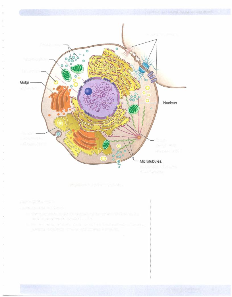
Ribosomes are composed of rRNA and protein. They consist of large (605) and small (405) subunits. Ribosomes are assembled in the nucleus and transported to the cytoplasm through the nuclear pores. The large ribosomal subunits are synthesized in the nucleolus, whereas the small subunits are syn thesized in the nucleus.
• Polysomes-Ribosomes often form polysomes, which consist of a single messenger RNA (mRNA) that is being translated by several ribosomes at the same time. The ribosomes move on the mRNA from the 5' end toward the 3' end. The 2 ribosomal subunits associate on the mRNA, with the small subunit binding first.
4MEDICAL
Section I • Histology and Cell Biology
Endoplasmic Reticulum
The endoplasmic reticulum exists in 2 forms, rough endoplasmic reticulum
(RER) and smooth endoplasmic reticulum (SER).
Rough endoplasmic reticulum
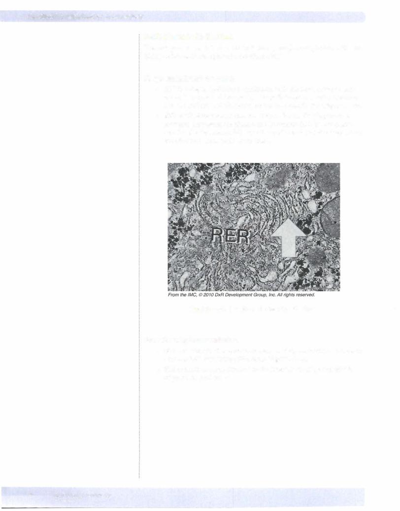
•RER is a single, lipid bilayer continuous with the outer nuclear mem brane. It is organized into stacks oflarge flattened sacs called cisternae that are studded with ribosomes on the cytoplasmic side (Figure I-1-3).
•RER synthesizes proteins that are destined for the Golgi apparatus, secretion, the plasma membrane, and lysosomes. RER is very promi nent in cells that are specialized in the synthesis of proteins destined for secretion (e.g., pancreatic acinar cells).
Figure 1-1-3. Rough endoplasmic reticulum
Smooth endoplasmic reticulum
•SER is a network of membranous sacs, vesicles, and tubules continuous with the RER, but lacking ribosomes (Figure 1-1-4).
•SER contains enzymes involved in the bio ynthesis of phospholipids, triglycerides, and sterols.
6MEDICAL