Материал: Kaplan USMLE-1 (2013) - Anatomy
Chapter i.
;
Copyright Lippincott Williams & Wilkins. Used with permission.
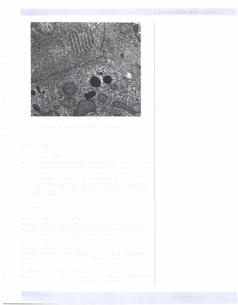
Figure 1-1-4. Human corpus luteum of pregnancy
Functions ofSER
Detoxification Reactions
These are reactions that make compounds water-soluble so that they can be excreted. Two types of reactions that increase solubility are:
•Hydroxylation reactions-by way of hydroxylase complexes containing cytochrome P450, a flavoprotein, and a nonheme iron protein
•Conjugation reactions-the transfer of polar groups (i.e., glucuronic
acid) from the active carrier UDP-glucuronic acid to the toxic water insoluble molecule
Steroid Synthesis
Glycogen Degradation and Gluconeogenesis
Removal of the phosphate group from glucose-6-phosphate by the enzyme glu cose-6 phosphatase, an integral membrane protein of the SER. This controls the formation of free glucose from glycogen and via gluconeogenesis.
Reactions in Lipid Metabolism
Lipolysis begins in the SER with the release of a fatty acid from triglyceride. The SER is also the site where lipoprotein particles are assembled.
Sequestration and Release of Calcium Ions
In striated muscle the SER is known as the sarcoplasmic reticulum (SR). The sequestration and release of calcium ions takes place in the SR.
• Cell Biology and Epithelia
MEDICAL 7
Section I • Histology and Cell Biology
Note |
Golgi Apparatus |
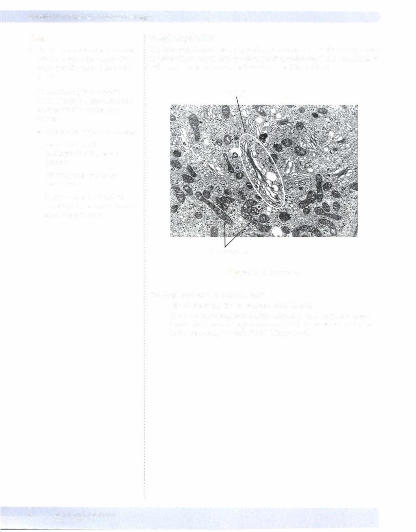
•Do not confuse the Golgi apparatus The Golgi apparatus consists ofdisc-shaped smooth cisternae that are assembled with the Golgi tendon organs of the in stacks (dictyosomes), having a diameter ofapproximately 1 µm and associated cell or any other factor bearing this with numerous small membrane-bound vesicles (Figure 1-1-5).
name.
• Dr. Camillo Golgi was a prolific |
Golgi |
Italian histologist. Other structures |
|
or processes bearing his name |
|
include: |
|
Golgi's silver stain for nerve cells |
|
- Cycle of Golgi for the |
|
development of the malaria |
|
parasite |
|
- Inhibitory Golgi cells of the |
|
cerebellum |
|
- Acroblast, a part of the Golgi |
|
material of the spermatid known |
|
as the Golgi remnant |
|
Mitochondria
Figure 1-1-5. Cytoplasm
The Golgi apparatus has 2 distinct faces:
•The cis (forming) face is associated with the RER.
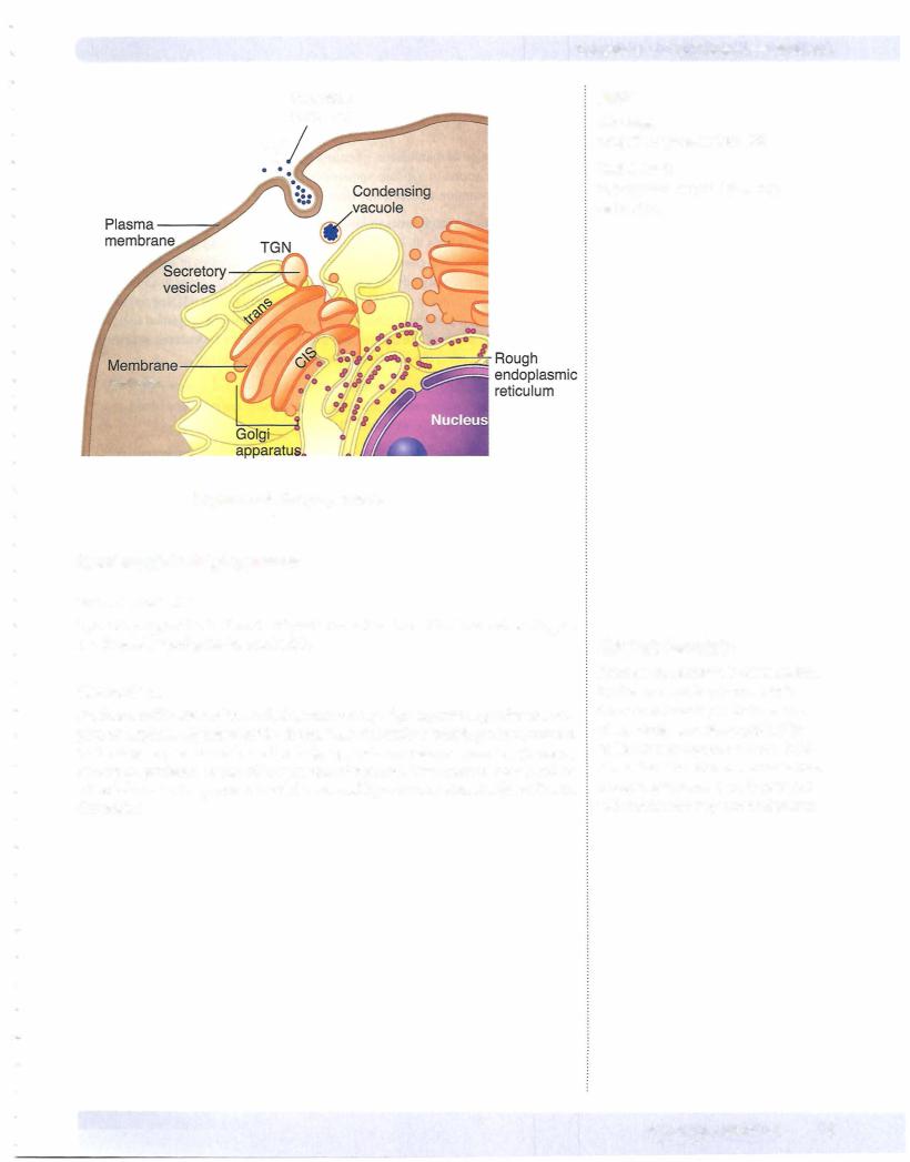
•The trans (maturing) face is often oriented toward the plasma mem brane. The trans-most region is a network of tubular structures known as the trans-Golgi network (TGN) (Figure I-1-6).
8MEDICAL
Section I • Histology and Cell Biology
Note
Lysosomal enzymes breakdown sphingolipids and glycoproteins into soluble products.
Clinical Correlate
I-Cell Disease
Phosphorylation of mannose in glycoproteins targets proteins to lysosomes. Phosphate is added in a 2-step sequence of reactions that are catalyzed by N-acetylglucosamine-phosphotransferase and N-acetylglucosaminidases.
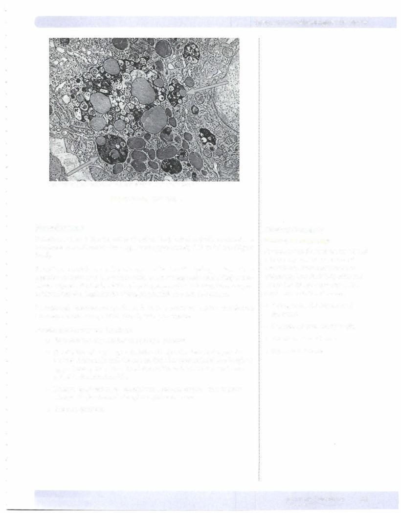
A deficiency in N-acetylglucosamine-phosphotransferase results in I-cell disease (mucolipidosis II), in which a whole family of enzymes is sent to the wrong destination. It is characterized by huge inclusion bodies in cells caused by the accumulation of undegraded glycoconjugates in lysosomes missing the hydrolases that normally degrade these macromolecules. The missing enzymes are found in the plasma and other body fluids, where they have normal levels of activity. The absence of the mannose-6-phosphate on the hydrolases results in their secretion rather than their incorporation into lysosomes.
The disease results in skeletal abnormalities, coarse features, restricted joint movements, and psychomotor retardation. Symptoms are generally noted at birth, and the life span is less than 10 years.
A somewhat less severe form of the disease with a later onset and potential survival into adulthood is called pseudo-Hurler polydystrophy.
There is no treatment for either disease, but prenatal diagnosis is available.
Clinical Correlate
Deficient breakdown of sphingolipids is cause of Gaucher, Niemann-Pick, and Tay-Sachs disease.
Lysosomes
Lysosomes are spherical membrane-enclosed organelles that are approximately 0.5 µm in diameter and contain enzymes required for intracellular digestion (Figure 1-1-7).
Lysosomes consist of 2 forms:
•Primarylysosomes have not yet acquired the materials to be digested. They are formed by budding from the trans side of the Golgi apparatus.
•Secondary lysosomes are formed by the fusion of the primary lysosome with the substrate to be degraded and have contents that are in various stages of degradation.
Lysosomes contain approximately 60 hydrolytic enzymes. These include nucle ases for degrading DNA and RNA, lipases for degrading lipids, glycosidases for degrading glycoconjugates (glycoproteins, proteoglycans, and glycolipids), prote ases and peptidases for degrading proteins, and a variety of phosphatases.
•All lysosomal enzymes are acid hydrolases, with optimal activity at a pH of approximately 5.0.
•The synthesis of the lysosomal hydrolases occurs in the RER; the hydro lases are transferred to the Golgi apparatus, where they are modified and packaged into lysosomes.
10 MEDICAL