Материал: Kaplan USMLE-1 (2013) - Anatomy
Section I • Histology and Cell Biology
Clinical Correlate
Three maternally inherited mitochondrial diseases that affect males more than females:
1. Leber's optic neuropathy
2. Pearson Marrow-Pancreas syndrome
3. Male infertility
Mitochondria
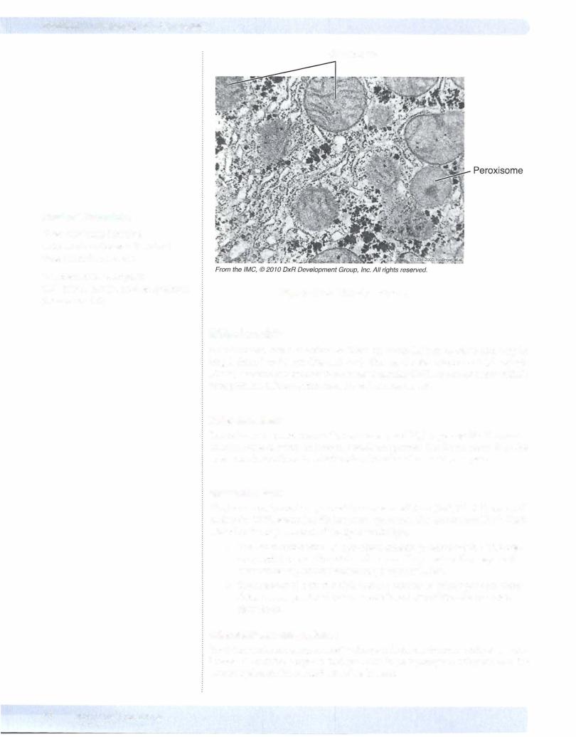
Figure 1-1 -8. The Peroxisome
Mitochondria
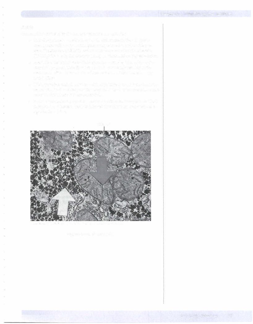
Mitochondria have 2 membranes. They are about 0.5 µ,m in width and vary in length from 1 to 10 µ,m (Figure I-1 -9). They synthesize adenosine triphosphate (ATP), contain their own double-stranded circular DNA, and make some oftheir own proteins. Mitochondria have several compartments.
Outermembrane
The outer membrane is smooth, continuous, and highly permeable. It contains an abundance of porin, an integral membrane protein that forms channels in the outer membrane through which molecules ofless than 10 kD can pass.
Innermembrane
The inner membrane is impermeable to most small ions (Na+, K+, H+) and small molecules (ATP, adenosine diphosphate, pyruvate). The impermeability is likely related to the high content of the lipid cardiolipin.
•The inner membrane has numerous infoldings, called cristae. The cris tae greatly increase the total surface area. They contain the enzymes for electron transportand oxidative phosphorylation.
•The number of mitochondria and the number of cristae per mitochon drion are proportional to the metabolic activity of the cells in which they reside.
lntermembrane compartment
The intermembrane compartment is the space between the inner and outer mem branes. It contains enzymes that use ATP to phosphorylate other nucleotides (creatine phosphokinase and adenylate kinase).
12 MEDICAL

Section I • Histology and Cell Biology
Clinical Correlate
A first step in the invasion of malignant cells through an epithelium results from a loss of expression of cadherins that weakens the epithelium.
Clinical Correlate
Changes in intermediate filaments are evident in neurons in Alzheimer's disease and in cirrhotic liver diseases.
Clinical Correlate
Colchicine prevents microtubule polymerization and is used to prevent neutrophil migration in gout. Vinblastine and vincristine are used in
cancer therapy because they inhibit the formation of the mitotic spindle.
CYTOSKELETAL ELEMENTS
Microfilaments
Microfilaments are actin proteins. They are composed of globular monomers of G-actin that polymerize to form helical filaments of F-actin. Actin polymeriza tion is ATP dependent. The F-actin filaments are 7-nm-diameter filaments that are constantly ongoing assembly and disassembly. F-actin has a distinct polarity. The barbed end (the plus end) is the site of polymerization and the pointed end is the site of depolymerization. Tread milling is the balance in the activity at the 2 ends.
In conjunction with myosin, actin microfilaments provide contractile and motile forces ofcells including the formation of a contractile ring that provides a basis for cytokinesis during mitosis and meiosis. Actin filaments are linked to cell mem branes at tight junctions and at the zonula adherens, and form the core ofmicrovilli.
Intermediate Filaments
Intermediate filaments are IO-nm-diameter filaments that are usually stable once formed. These filaments provide structural stability to cells. There are 4 groups of intermediate filaments:
•Type I is keratins. Keratins are found in allepithelial cells.
•Type II is intermediate filaments comprising a diverse group.
Desmin is found in skeletal, cardiac, and gastrointestinal (GI) tract smooth muscle cells.
Vimentin is found in most fibroblasts, fibrocytes, endothelial cells, and vascular smooth muscle.
Glial fibrillary acidic protein is found in astrocytes and some Schwann cells.
Peripherin is found in peripheral nerve axons.
•Type III is intermediate filaments forming neurofilaments in neurons.
•Type IV is 3 types of lamins which form a meshwork rather than indi vidual filaments inside the nuclear envelope of all cells.
Microtubules
Microtubules consist of 25-nm-diameter hollow tubes. Like actin, microtubules are undergoing continuous assembly and disassembly. They provide "tracks" for intracellular transport of vesicles and molecules. Such transport exists in all cells but is particularly important in axons. Transport requires specific ATPase motor molecules; dynein drives retrograde transport and kinesin drives anterograde transport. Microtubules are found in true cilia andflagella, and utilize dynein to convey motility to these structures. Microtubules form the mitotic spindle during mitosis and meiosis.
CELL ADHESION MOLECULES
Cell adhesion molecules are surface molecules that allow cells to adhere to one another or to components of the extracellular matrix. The expression ofadhesion molecules on the surface ofa given cell may change with time, altering its interac tion with adjacent cells or the extracellular matrix.
14 MEDICAL

Section I • Histology and Cell Biology
Clinical Correlate
Pemphigus Vulgaris (autoantibodies against desmosomal proteins in skin cells)
•Painful flaccid bullae (blisters) in oropharynx and skin that rupture easily
•Postinflammatory hyperpigmentation
•Treatment: corticosteroids
Bullous Pemphigoid (autoantibodies against basement-membrane hemidesmosomal proteins)
•Widespread blistering with pruritus
•Less severe than pemphigus vulgaris
•Rarely affects oral mucosa
•Can be drug-induced (e.g., middle aged or elderly patient on multiple medications)
•Treatment: corticosteroids
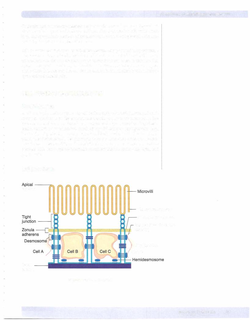
Tight junctions (zonula occludens) function as barriers to diffusion and deter mine cell polarity. They form a series of punctate contacts of adjacent epithelial cells near the apical end or luminal surface of epithelial cells. The major com ponents of tight junctions are occludins (Z0-1,2,3) and claudin proteins. These proteins span between the adjacent cell membranes and their cytoplasmic parts bind to actin microfilaments.
Zonula adherens forms a belt around the entire apicolateral circumference of the cell, immediately below the tight junction of epithelium. Cadherins span between the cell membranes. Like the tight junctions immediately above them, the cyto plasmic parts of cadherins are associated with actin filaments.
Desmosomes (maculaadherens) function as anchoring junctions. Desmosomes provide a structural and mechanical link between cells. Cadherins span between the cell membranes of desmosomes and internally desmosomes are anchored to intermediate filaments in large bundles called tonofilarnents.
Hemidesmosomes adhere epithelial cells to the basement membrane. The basement membrane is a structure that consists of the basal membrane of a cell and 2 underlying extracellular components, the basal lamina and the reticular lamina. The basal lamina is a thin felt-like extracellular layer composed of pre dominantly of type IV collagen associated with laminin, proteoglycans, and fibronectin that are secreted by epithelial cells. Fibronectin binds to integrins on the cell membrane, and fibronectin and laminin in turn bind to collagen in the basal lamina. Internally, like a desmosome, the hemidesmosomes are linked to intermediate filaments. Below the basal lamina is the reticular lamina, com posed of reticular fibers.
Through the binding of extracellular components of hemidesmosomes to inte grins, and thus to fibronectin and laminin, the cell is attached to the basement membrane and therefore to the extracellular matrix components outside the basement membrane. These interactions between the cell cytoplasm and the extracellular matrix have implications for permeability, cell motility during em bryogenesis, and cell invasion by malignant neoplasms.
Gap junctions (communicating junctions) function in cell-to-cell communica tion between the cytoplasm of adjacent cells by providing a passageway for ions such as calcium and small molecules such as cyclic adenosine monophosphate (cAMP). The transcellular channels that make up a gap junction consist of con nexons, which are hollow channels spanning the plasma membrane. Each con nexon consists of 6 connexin molecules. Unlike other intercellular junctions, gap junctions are not associated with any cytoskeletal filament.
16 MEDICAL