Section Ill • Gross Anatomy
Truncus arteriosus defects
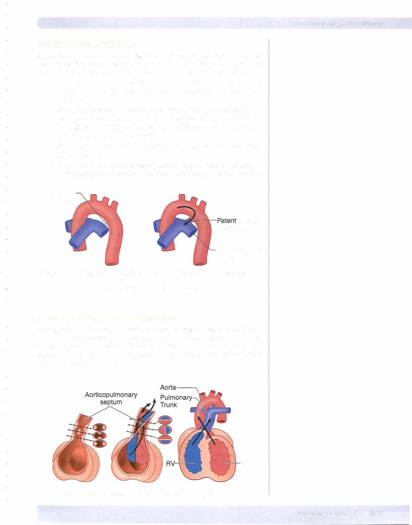
Three classic cyanotic congenital heart abnormalities occur with defects in the development of the aorticopulmonary septum and are related to the failure of neural crest cells to migrate into the truncus arteriosus:
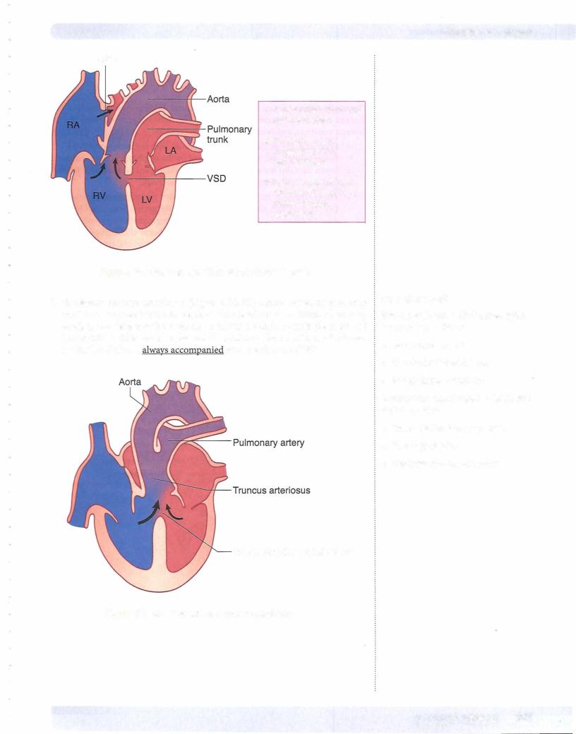
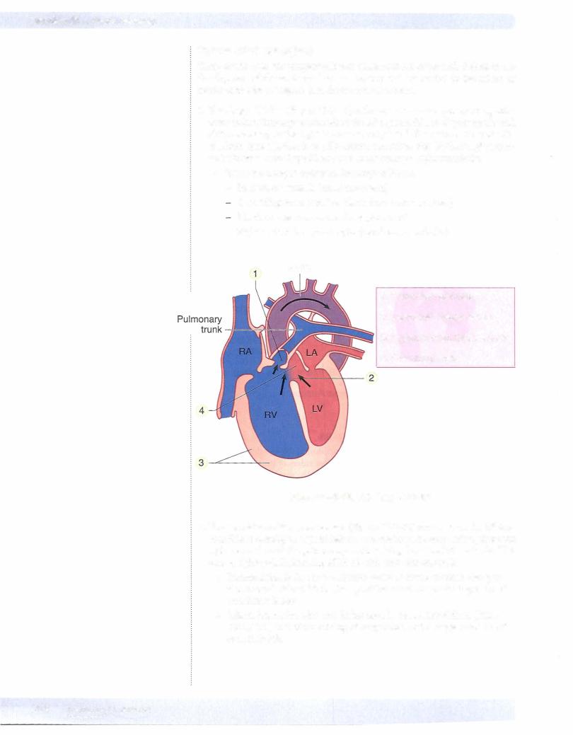
1 . TetralogyofFallot (Figure III-2- 1 9) is the mostcommon cyanotic congenital heart defect. Tetralogy occurs when the AP septum fails to align properly and shifts anteriorly to the right. This causes right-to-left shunting of blood with resultant cyanosis that is usually present sometime after birth. Imaging typi cally shows a boot-shaped heart due to the enlarged right ventricule.
•There are 4 major defects in Tetralogy of Fallot:
-Pulmonary stenosis (most important)
Overriding aorta (receives blood from both ventricles) Membranous interventricular septal defect
-Right ventricular hypertrophy (develops secondarily)
Aorta
1 . Pulmonary stenosis
2. Ventricular septa! defect
3. Hypertrophied right ventricle
4. Overriding aorta
Figure 111-2-19. Tetralogy of Fallot
2.Transposition ofthe great vessels (Figure Ill-2-20) occurs when the AP sep tum fails to develop in a spiral fashion and results in the aorta arising from the right ventricle and the pulmonary trunk arising from the left ventricle. This causes right-to-left shunting of blood with resultant cyanosis.
•Transposition is the most common cause of severe cyanosis that per sists immediately at birth. Transposition results in producing 2 closed circulation loops.
•Infants born alive with this defect usually have other defects (PDA, VSD, ASD) that allow mixing of oxygenated and deoxygenated blood to sustain life.