Материал: Kaplan USMLE-1 (2013) - Anatomy

Внимание! Если размещение файла нарушает Ваши авторские права, то обязательно сообщите нам

Chapter 2 • Thorax

Anterior Mediastinum

The anterior mediastinum is the small interval between the sternum and the anterior surface of the pericardium. It contains fat and areolar tissue and the infe­ rior part ofthe thymus gland. A tumor ofthe thymus (thymoma) can develop in the anterior or superior mediastina

Posterior Mediastinum

Theposteriormediastinumis located between the posterior surface ofthe pericar­ dium and the T5-Tl2 thoracic vertebrae. Inferiorly, it is closed by the diaphragm.

There are 4 vertically oriented structures coursing within the posterior medias­ tinum:

•Thoracic (descending) aorta

-Important branches are the bronchial, esophageal, and posterior inter­ costal arteries

-Passes through the aortic hiatus (with the thoracic duct) at the T12 vertebral level to become the abdominal aorta

•Esophagus

-Lies immediately posterior to the left primary bronchus and the left atrium, forming an important radiological relationship

-

Covered by the anterior and posterior esophagealplexuseswhich are

-

Passes through the esophageal hiatus (with the vagal nerve trunks) at


derived from the left and right vagus nerves, respectively


the Tl0 vertebral level

-Is constricted (1) at its origin from the pharynx, (2) posterior to the arch of the aorta, (3) posterior to the left primary bronchus, and (4) at the esophageal hiatus of the diaphragm.

•Thoracic duct

-Lies posterior to the esophagus and between the thoracic aorta and azygos vein

-Ascends the posterior and superior mediastina and drains into the junction of the left subclavian and internal jugular veins

-Arises from the cisterna chyli in the abdomen (at vertebral level L1) and enters the mediastinum through the aortic hiatus of the diaphragm

•Azygos system ofveins

-Drains the posterior and thoracic lateral wall

-Communicates with the inferior vena cava in the abdomen and ter- minates by arching over the root of the right lung to empty into the superiorvena cava above the pericardium

-Forms a collateralvenous circulation between the inferior and superior vena cava

Middle Mediastinum

The middle mediastinum contains the heart and great vessels and pericardium and willbe discussed later.
MEDICAL 207

111-2-23).

Chapter 2 • Thorax

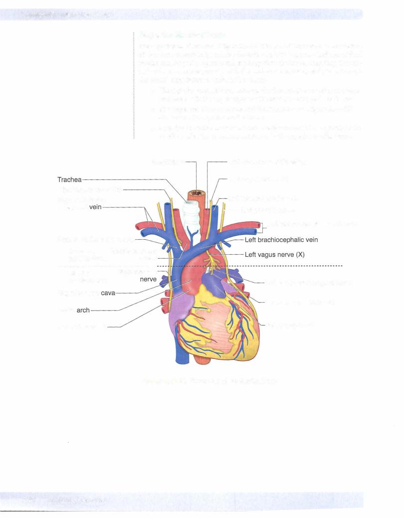
The relationships ofthese structures in the superior mediastinum are best visual­ ized in a ventral to dorsal orientation between the sternum anteriorly and the vertebrae posteriorly:

• Thymus: Located posterior to the manubrium, usually atrophies in the adult and remains as fatty tissue

• Rightandleftbrachiocephalicveins: Rightvein descends almostvertically and the left vein obliquely crosses the superiormediastinumposterior to the thyrnic remnants.

The 2 veins join to form the superiorvenacava posterior to the right first costalcartilage.

The superiorvena cava descends and drains into the right atrium deep to the right third costal cartilage.

• Aortic arch and its 3 branches: Aortic arch begins and ends at the plane ofthe sternal angle and is located just inferior to the left brachiocephalic vein. As a very important radiological landmark, the origins ofthe

3 branches ofthe aortic arch (brachiocephalic,left common carotid,and left subclavian) are directly posterior to the left brachiocephalic vein.

• Trachea: Lies posterior to the aortic arch and bifurcates at the level of

T4 vertebra to form the right and left primary bronchi. The carina is an internal projection of cartilage at the bifurcation.

• Esophagus: Lies posterior to the trachea and courses posterior to the left primary bronchus to enter the posterior mediastinum

ln addition to these structures, the superior mediastinum also contains the right and leftvagus and phrenic nerves and the superior end ofthe thoracic duct.

Vogus nerves

•Right and left vagus nerves contribute to the pulmonary and cardiac plexuses.

•In the neck, the right vagus nerve gives rise to the right recurrent laryn­ geal nerve, which passes under the right sublcavian artery to ascend in the groove between the esophagus and the trachea to reach the larynx. Note: The right recurrent laryngeal nerve is not in the mediastinum.

•The left vagus nerve gives rise to the left recurrent laryngealnervein

the superior mediastinum, which passes under the aortic arch and liga­ mentum arteriosum to ascend to the larynx (Figure

Thoracic duct

The thoracic duct is the largest lymphatic channel in the body. It returns lymph to the venous circulation at the junction ofthe left internal jugular vein and the left subclavian vein.

Phrenicnerves

Phrenic nerves arise from the ventral rami of cervical nerves 3, 4, and 5. The nerves are the sole motor supply of the diaphragm and convey sensory infor­ mation from the central portion of both the superior and inferior portions of the diaphragm and parietal pleura. Both phrenic nerves pass through the middle mediatstinum lateral between the fibrous pericardium and pleura, and anterior to the root ofthe lung.

Clinical Correlate

The left recurrent laryngeal nerve

(Figure 111-2-23) curves under the aortic arch distal to the ligamentum arteriosum where it may be damaged by pathology (e.g., malignancy or aneurysm of the aortic arch), resulting in paralysis ofthe leftvocal folds. The right laryngeal nerve is not affected because it arises from the right vagus

nerve in the root ofthe neck and passes under the subclavian artery.

Either the right or the left recurrent laryngeal nerve may be lesioned with thyroid gland surgery.

MEDICAL 209

Источник: https://studfile.net/preview/14638320/