Материал: Kaplan USMLE-1 (2013) - Anatomy

Внимание! Если размещение файла нарушает Ваши авторские права, то обязательно сообщите нам
right-to-leftpres­
ductus arteriosus

Chapter 2 • Thorax

Fetal Circulation

Venoussystems associated with thefetal heart

There are 3 major venous systems that flow into the sinus venosus end of the heart tube.

• Viteline (omphalomesenteric) veins drain deoxygenated blood from the yolk stalk; they will coalesce and form the veins of the liver (sinusoids, hepatic portal vein, hepatic vein) and part of the inferior vena cava.

•Umbilicalvein carries oxygenated blood from the placenta.

•Cardinalveins carry deoxygenated blood from the body of the embryo;

they will coalesce and contribute to some of the major veins of the body (brachiocephalic, superior vena cave, inferior vena cava, azygos, renal).

Arterialsystems associated with thefetal heart

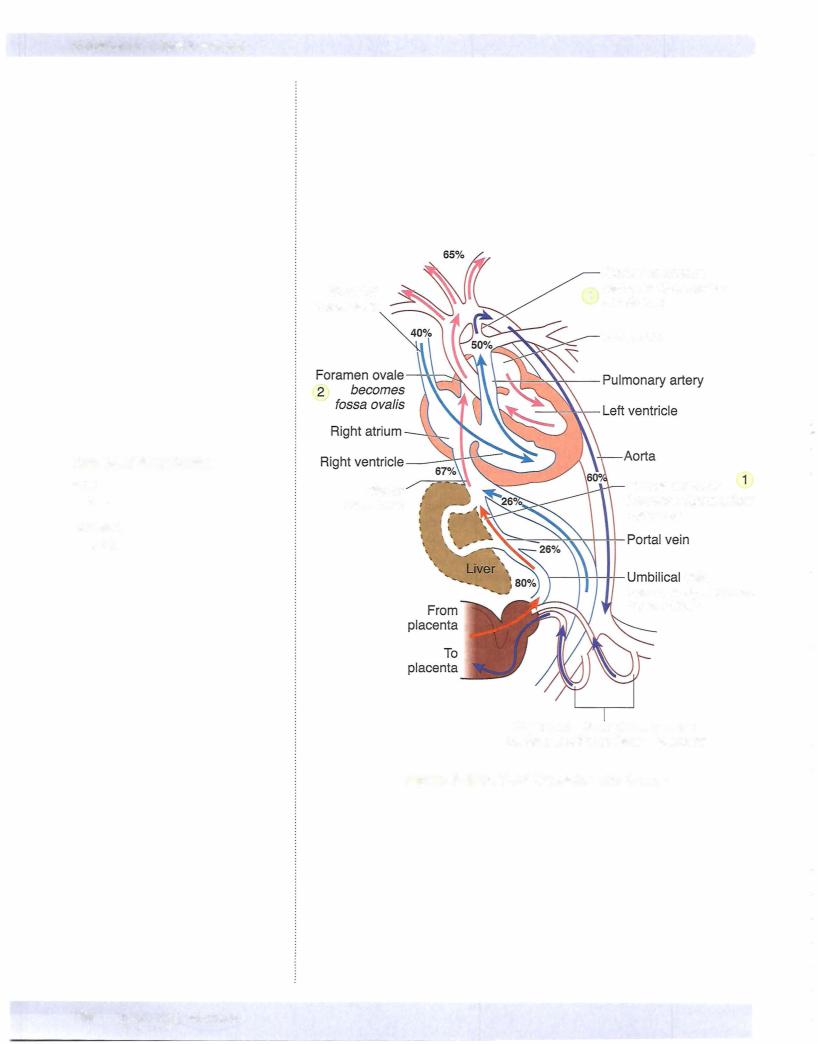
During fetal circulation, oxygenated blood flood from the placenta to the fetus passes through the umbilical vein. Three vascular shunts develop in the fetal circulation to bypass blood flow around the liver and l gs (Figure IIl-2-12).

1 . The ductus venosus allows oxygenated blood in the umbilical vein to bypass the sinusoids of the liver into the inferior vena cava and to the right atrium. From the right atrium, oxygenated blood flows mostly through the foramen ovale into the left atrium then left ventricle and into the systemic circulation.

2. The foramen ovale develops during atrial septation to allow oxygenated blood to bypass the pulmonary circulation. Note that this is a right-to­ left shunting of blood during fetal life.

3. During fetal circulation, the superior vena cava drains deoxygenated blood from the upper limbs and head into the right atrium. Most of this blood flow is directed into the right ventricle and into the pulmonary trunk. The opens into the underside of the aorta just distal to the origin of the left subclavian artery and shunts this deoxy­ genated blood from the pulmonary trunk to the aorta to bypass the pul­ monary circulation.

The shunting of blood through the foramen ovale and through the ductus arteriosus (right to left) during fetal life occurs because of a

sure gradient.

MEDICAL 197

Chapter 2 • Thorax

Postnatal circulation

Following birth, these 3 shunts will close because of changes in the pressure gra­ dients and in oxygen tensions. The umbilical vein closes and reduces blood flow into the right atrium. The ductus venosus also closes. Lung expansion reduces pulmonary resistence and results in increased flow to the lungs and increased venous return to the left atrium.

•Closure of the foramen ovale occurs as a result of the increase in left atrial pressure and reduction in right atrial pressure.

•Closure of the ductus venosus and ductus arteriosus occurs over the next several hours as a result of the contraction of smooth muscles in its wall and increased oxygen tension.

•The release of bradykinin and the immediate drop of prostaglandin E at birth also facilitate the closure of the ductus arteriosus.

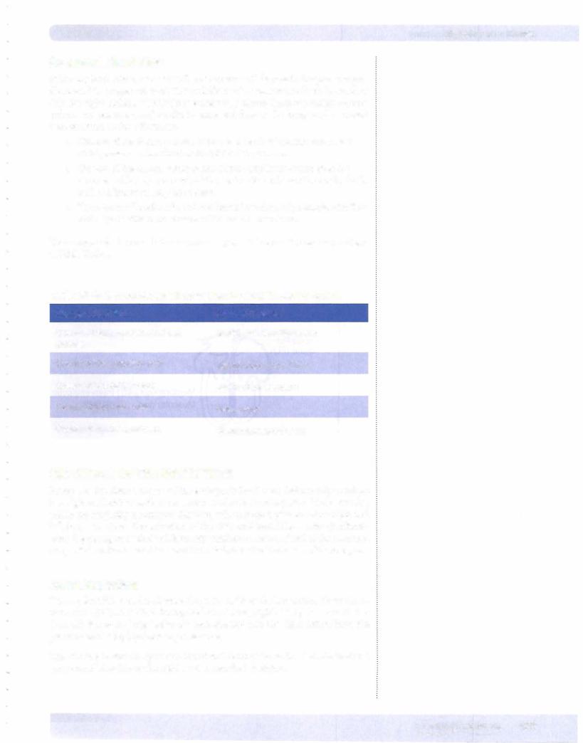
The changes which occur between preand postnatal circulation are summarized in Table ill-2-2.

Table 111-2-2. AdultVestiges Derived from the Fetal Circulatory System

Changes After Birth

Remnant in Adult

Closure of right and left umbilical

Medial umbilical ligaments

arteries


Closure ofthe umbilical vein

Ligamentum teres of liver

Closure of ductus venosus

Ligamentum venosum

Closure of foramen ovale

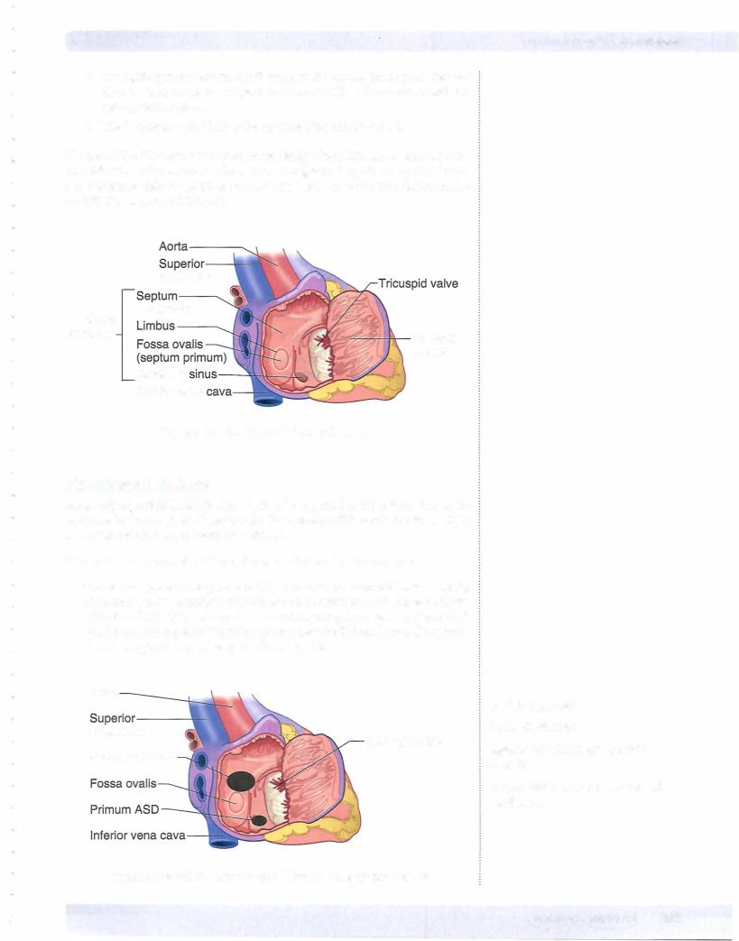
Fossa ovalis

Closure of ductus arteriosus

Ligamentum arteriosum

SEPTATION OF THE HEART TUBE

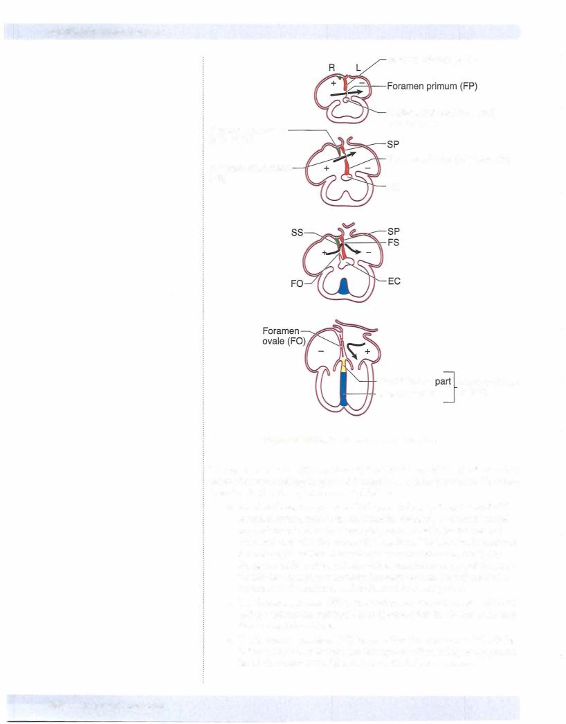
Except for the sinus venosus of the embryonic heart tube that initially develops into right and left horns, the ventricular, atrial, and truncus parts ofthe heart tube, which are originally a common chamber, willundergo septation into a right and left heart structure. The septation of the atria and ventricles occurs simultane­ ously beginning in week 4 and is mostly finished in week 8. Most ofthe common congenital cardiac anomalies result from defects in the formation ofthese septa.

Atrial Septation

During fetal life, blood is shunted from the right to the left atrium via the fora­ men ovale (FO). Note that during fetal circulation, right atrial pressure is higher than left due to the large bolus of blood directed into the right atrium from the placenta and to high pulmonary resistance.

The FO has to remain open and functional during the entire fetal life to shunt oxygenated blood from the right atrium into the left atrium.

MEDICAL 199

Источник: https://studfile.net/preview/14638320/