Материал: Kaplan USMLE-1 (2013) - Anatomy

Внимание! Если размещение файла нарушает Ваши авторские права, то обязательно сообщите нам

Section Ill • Gross Anatomy

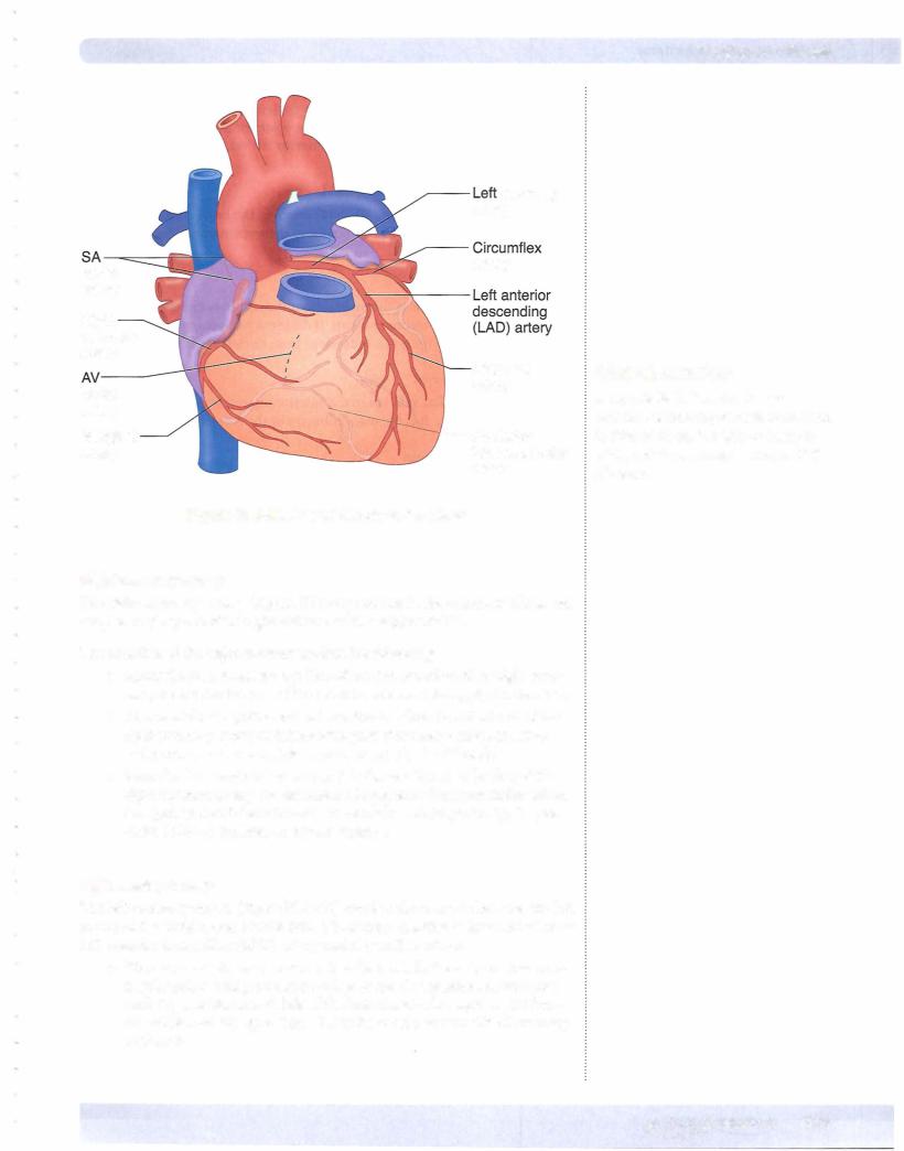
•The circumflex artery courses around the left border of the heart in the coronary sulcus and supplies ( 1) the left border ofthe heart via the marginal branch and (2) ends on the posterior aspect of the left ven­ tricle and supplies the posterior-inferior left ventricular wall.

Venous Drainage ofthe Heart

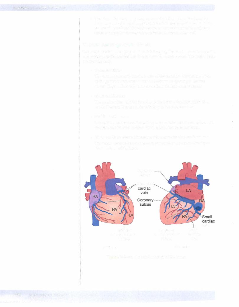
The major cardiac veins (Figure IIl-2-34) draining the heart course in the sulci and accompany the arteries but do not carry the same names. The major veins are the following:

•Coronarysinus

The coronary sinus is the main vein of the coronary circulation; it lies in the posterior coronary sulcus. It drains to an opening in the right atrium (Figure III-2-34). It develops from the left sinus venosus.

•Great cardiacvein

The great cardiac vein lies in the anterior interventricular sulcus with the LAD artery. It is the main tributary of the coronary sinus.

•Middle cardiac vein

The middle cardiac vein lies in the posterior interventricular sulcus with the posterior interventricular artery. It joins the coronary sinus.

•Venae cordis minimae (thebesian veins) and anterior cardiac veins

The venae cordis minimae and anterior cardiac veins open directly to the chambers of the heart.

Coronary

sinus

Great

vein

Anterior

Posterior

Middle

interventricular

interventricular

cardiac

sulcus

sulcus

vein

Anterior

Posterior

Figure 111-2-34. Venous Drainage of the Heart

220 MEDICAL

Chapter 2 • Thorax

Conducting System ofthe Heart

SA node

The SA node initiates the impulse for contraction ofheart muscle (and is therefore termed the "pacemaker" of the heart). It is located at the superior end of the crista terminalis, where the superior vena cava enters the right atrium (Figure IIl-2-35).

The SA node is supplied bythe SA nodal branch ofthe right coronary artery.

Impulse production is speeded up by sympathetic nervous stimulation; it is slowed by parasympathetic (vagal) stimulation.

AVnode

The AV node receives impulses from the SA node. The AV node is located in the interatrial septum near the opening ofthe coronary sinus. The AV node slows the impulse so that it reaches the ventricles after it has reached the atria.

The AV node is supplied by the rightcoronaryartery.

Bundle ofHis

The bundle ofHis originates in the AV node. It conducts impulses to the right and left ventricles. It is supplied by the LAD artery.

In the right ventricle, the moderator band (septomarginal trabecula) contains the right bundle branch.

Impulses pass from the right and left bundle branches to the papillary muscles and ventricular myocardium.

Innervation

The cardiac plexus is a combination of sympathetic and parasympathetic (vagal) fibers.

•Sympathetic stimulation increases the heart rate. Nerves that sense pain associated with coronary artery ischemia (angina) follow the sympa­ thetic pathways back into spinal cord segments Tl-TS.

•Parasympathetic stimulation slows the heart rate. Sensory nerves that carry the afferent limb of cardiac reflexes travel with the vagus nerve.
MEDICAL 221

Источник: https://studfile.net/preview/14638320/