Section Ill • Gross Anatomy
Clinical Correlate
Spastic bladder results from lesions of the spinal cord above the sacral spinal cord levels. There is a loss of inhibition ofthe parasympathetic nerve fibers that innervate the detrusor muscle during the filling stage. Thus, the detrusor muscle responds to a minimum amount of stretch, causing urge incontinence.
Clinical Correlate
Atonic bladder results from lesions to the sacral spinal cord segments or the sacral spinal nerve roots. Loss of pelvic splanchnic motor innervation
with loss of contraction of the detrusor muscle results in a full bladder with a continuous dribble of urine from the bladder.
Clinical Correlate
Weakness of the puborectalis part of the levator ani muscle may result in rectal incontinence.
Weakness of the sphincter urethrae part of the urogenital diaphragm may result in urinary incontinence.
268 MEDICAL
• Muscles
The detrusor muscle forms most of the smooth-muscle walls of the bladder and contracts during emptying of the bladder (micturition). The contraction of these muscles is under control of the parasympa thetic fibers of the pelvic splanchnics (52, 3, 4)
The internal urethral sphincter (sphincter vesicae) is smooth-muscle fibers that enclose the origin of the urethra at the neck ofthe bladder. These muscles are under control of the sympathetic fibers of the lower thoracic and lumbar splanchnics (Tl l -L2) and are activated during the filling phase of the bladder to prevent urinary leakage.
-The external urethralsphincter (sphincter urethrae) is the voluntary skeletal muscle component of the urogenital diaphragm that encloses the urethra and is relaxed during micturition (voluntary muscle
of micturition). The external sphincter is innervated by perineal branches of the pudendal nerve.
Urethra
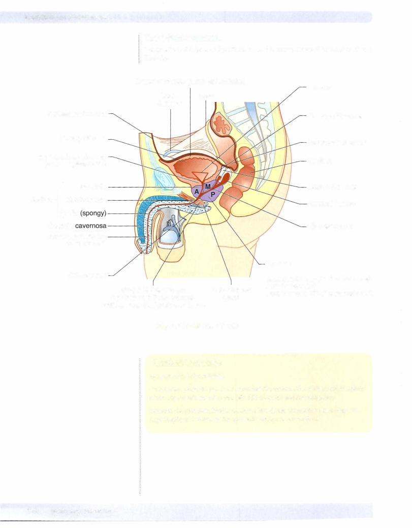
The male urethra is a muscular tube approximately 20 cm in length. The urethra in men extends from the neck of the bladder through the prostate gland (prostat ic urethra) to the urogenital diaphragm ofthe perineum (membranous urethra), and then to the external opening of the glans (penile or spongy urethra) (Figure
III-3-26).
The male urethra is anatomically divided into 3 portions: prostatic, membra nous, and spongy (penile).
•The distal spongyurethra of the male is derived from the ectodermal cells of the glans penis.
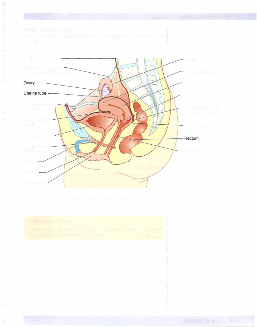
The female urethra is approximately 4 cm in length and extends from the neck of the bladder to the external urethral orifice ofthe vulva (Figure III-3-27).
PELVIS
Pelvic and Urogenital Diaphragms
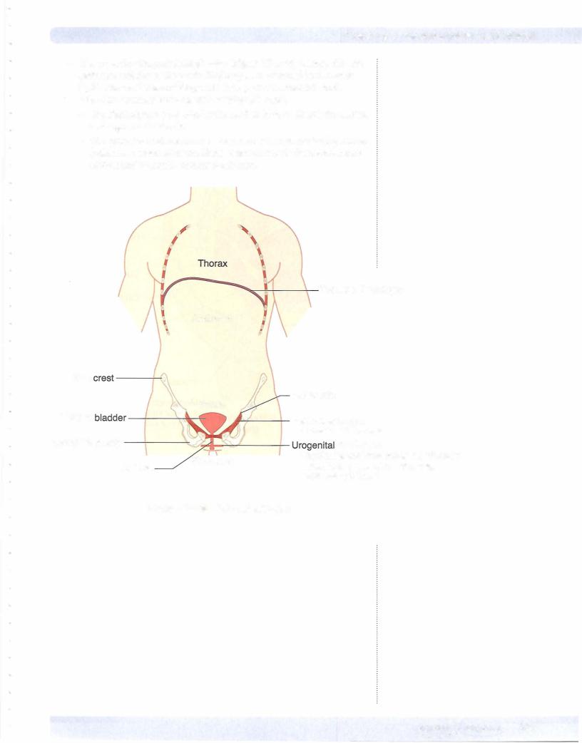
The pelvic andurogenital diaphragms (Figure III-3-25) are 2 important skeletal muscle diaphragms that provide support of the pelvic and perinea! structures. They are each innervated by branches of the pudenda! nerve.
•The pelvic diaphragm forms the muscular floor of the pelvis and separates the pelvic cavity from the perineum. The pelvic diaphragm is a strong support for the pelvic organs and transmits the distal parts of the genitourinary system and GI tract from the pelvis to the perineum.
-The diaphragm is formed by 2 layers of fascia and the 2 muscles: the levator ani and coccygeus.
-The puborectalis component of the levator ani muscle forms a muscu
lar sling around the anorectal junction, marks the boundary between the rectum and anal canal and is important in fecal continence.