Clinical Correlate
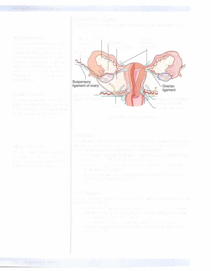
The ureter passes inferior to the uterine artery 1 to 2 centimeters from the cervix ("water under the bridge") and must be avoided during surgical procedures.
Clinical Correlate
A pudenda( nerve block to anesthetize the perineum is performed as the pudenda! nerve crosses posterior to the ischial spine (Figure 111-3-248).
Uterine artery |
|
Transverse (cardinal) |
("water under bridge") |
|
|
Ureter |
cervical ligament |
|
Uterosacral ligament |
|
|
|
Figure 111-3-28. Broad Ligament |
|
PERINEUM
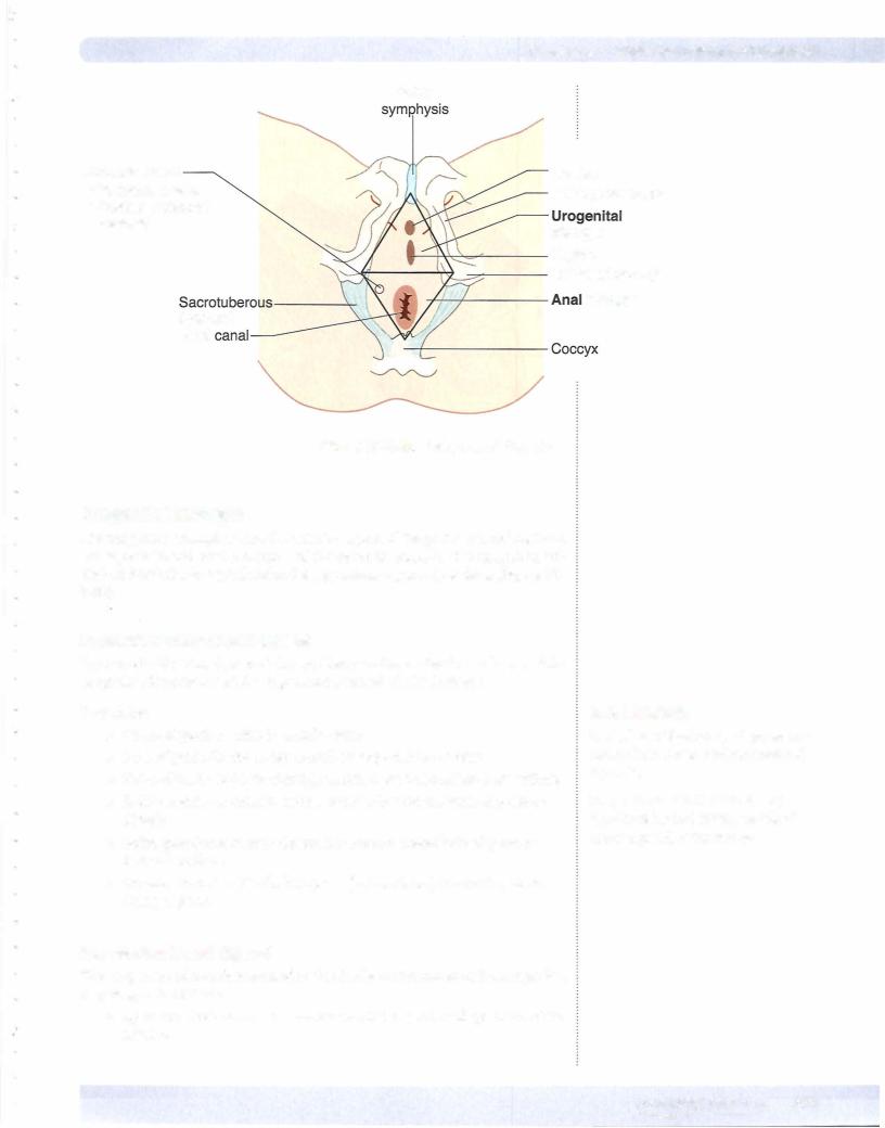
The perineum is the diamond-shaped outlet of the pelvis located below the pelvic diaphragm. The perineum is divided by a transverse line between the ischial tuber osities into the anal and urogenital triangles (Figure III-3-29).
•The sensory and motor innervation to the perineum is provided by the pudenda! nerve (S2, 3, 4) of the sacral plexus.
•The blood supply is provided by the internal pudenda! artery, a branch of the internal iliac artery.
•The pudendal nerve and vessels cross the ischial spine posteriorly to enter the perineum (Figure III-3-24 B).
AnalTriangle
The anal triangle is posterior and contains the anal canal surrounded by the fat-filled ischioanal fossa.
•The anal canal is guarded by a smooth-muscle internal anal sphincter innervated by the ANS and an externalanal sphincter of skeletal mus cle innervated by the pudendal nerve.
•The pudenda! canal transmitting the pudenda! nerve and internal pudenda! vessels is found on the lateral aspect of the ischioanal fossa (Figure III-3-29) .