Материал: Kaplan USMLE-1 (2013) - Anatomy
Внимание! Если размещение файла нарушает Ваши авторские права, то обязательно сообщите нам

Chapter 3 • Abdomen, Pelvis, and Perineum

POSTERIOR ABDOMINAL BODYWALL

Embryology of Kidneys and Ureter

Renal development is characterized by 3 successive, slightly overlapping kidney systems: pronephros, mesonephros, and metanephros (Figure IIl-3-21).

Stomach

Midgut

Cecum

Pronephros

Mesonephros

Hindgut

Figure 111-3-21 . Pronephros, Mesonephros, and Metanephros

Pronephros

During week 4, segmented nephrotomes appear in the cervical intermediate me­ soderm ofthe embryo. These structures grow laterally and canalize to form neph­ ric tubules. The first tubules formed regress before the last ones are formed. By the end ofweek 4, the pronephros disappears and does not function.

Mesonephros

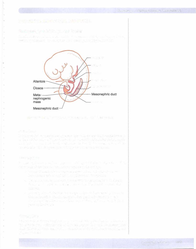
In week 5, the mesonephros appears as S-shaped tubules in the intermediate mesoderm of the thoracic and lumbar regions of the embryo.

•The medial end of each tubule enlarges to form a Bowman's capsule into which a tuft of capillaries, or glomerulus, invaginates.

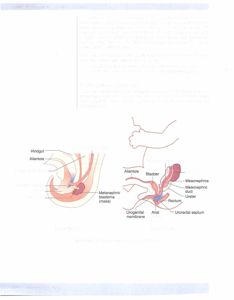
•The lateral end of each tubule opens into the mesonephric (Wolffian) duct, an intermediate mesoderm derivative. The duct drains into the hindgut.

•Mesonephric tubules function temporarily and degenerate by the begin­ ning of month 3. The mesonephric duct persists in the male as the ductus epididyrnidis, ductus deferens, and the ejaculatory duct. It disap­ pears in the female.

Metanephros

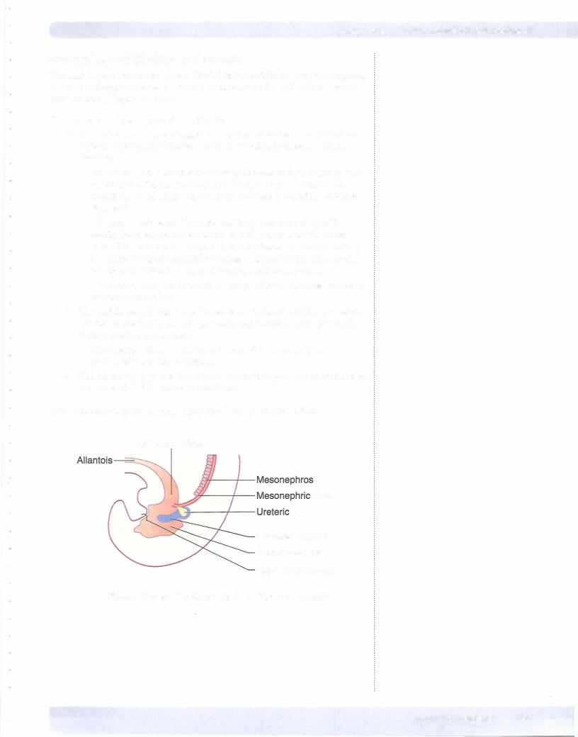
During week 5, the metanephros, or permanent kidney, develops from 2 sources: the uretericbud, a diverticulum of the mesonephric duct, and the metanephric mass (blastema), from intermediate mesoderm of the lumbar and sacral regions (Figure IIl-3-22).

MEDICAL 263
Section Ill • Gross Anatomy

Clinical Correlate

Blockage by Renal Calculi

The most common sites of ureteral constriction susceptible to blockage by renal calculi are:

•Where the renal pelvis joins the ureter

•Where the ureter crosses the pelvic inlet

•Where the ureter enters the wall of the urinary bladder

Congenital Abnormalities ofthe Renal System

•Renal agenesis

Renal agenesis results from failure of one or both kidneys to develop because of early degeneration of the ureteric bud. Unilateral genesis is fairly common; bilateral agenesis is fatal (associated with oligohydram­ nios, and the fetus may have Pottersequence: clubbed feet, pulmonary hypoplasia, and craniofacial anomalies) .

•Pelvic and horseshoekidney

Pelvic kidney is caused by a failure of one kidney to ascend. Horseshoe kidney (usually normalrenalfunction, predisposition to calculi) is a fusion of both kidneys at their ends and failure of the fused kidney to ascend. The horseshoe kidney hooks under the origin of the inferior mesenteric artery.

•Double ureter

Caused by the early splitting of the ureteric bud or the development of 2 separate buds

•Patent urachus

Failure ofthe allantois to be obliterated results in urachal fistulas or sinuses. In male children with congenital valvular obstruction of the pros­ tatic urethra or in older men with enlarged prostates, a patent urachus maycause drainage ofurine through the umbilicus.

Posterior Abdominal Wall and Pelvic Viscera

Kidneys

The kidneys are a pair ofbean-shaped organs approximately 1 2 cm long. They ex­ tend from vertebral level Tl2 to L3 when the body is in the erect position (Figure III-3-24A and -24B). The right kidney is positioned slightly lower than the left because of the mass of the liver.

•Kidney's Relation to the PosteriorAbdominalWall

Both kidneys are in contact with the diaphragm, psoas major, and qua­ dratus lumborum (Figure III-3-24) .

-Right kidney-contacts the above structures and the 1 2th rib

-Left kidney-contacts the above structures and the 1 1th and 12th ribs

Ureters

Ureters are fibromuscular tubes that connect the kidneys to the urinary bladder in the pelvis. They run posterior to the ductus deferens in males and posterior to the uterine artery in females. They begin as continuations of the renal pelves and run retroperitoneally, crossing the external iliac arteries as they pass over the pelvic brim.

Ureter's Relation to the PosteriorAbdominalWall

The ureter lies on the anterior surface of the psoas major muscle.

266 MEDICAL

Источник: https://studfile.net/preview/14638320/