Материал: Kaplan USMLE-1 (2013) - Anatomy

Внимание! Если размещение файла нарушает Ваши авторские права, то обязательно сообщите нам

Chapter 10 • Cerebral Cortex

Clinical Correlate (Continued)

Asomatognosia

The integration ofvisual and somatosensory information is important for the formation ofthe "body image" and awareness ofthe body and its position in space. Widespread lesions in areas 7, 39, and 40 in the nondominant right parietal lobe may result in unawareness or neglect of the contralateral half of the body, known as asomatognosia. Although somatic sensation is intact, the patients ignore half oftheir body and may fail to dress, undress, or wash the affected (left) side. Patients will have no visual field deficits, so they can see, but deny the existence ofthings in the left visual field. Asking them to bisect a horizontal line produces a point well to the right of true center. If asked to draw a clock face from memory, they will draw only the numbers on the right side, ignoring those on the left. Patients may deny that the left arm or leg belongs to them when the affected limb is passively brought into their field ofvision. Patients may also deny their deficit, an anosognosia.

Occipital Lobe

The occipital lobe is essential for the reception and recognition of visual stimuli and contains primary visual and visual association cortex.

Visual cortex

The visual cortex is divided into striate (area 17) and extrastriate (areas 18 and 19). Area 17, also referred to as the primary visual cortex, lies on the medial por­ tion ofthe occipital lobe on either side ofthe calcarine sulcus. Its major thalamic input is from the lateral geniculate nucleus. Some input fibers are gathered in a thick bundle that can be visible on the cut surface of the gross brain, called the line of Gennari. The retinal surface (and therefore the visual field) is represented in an orderly manner on the surface of area 17, such that damage to a discrete part of area 17 willproduce a scotoma (i.e., a blind spot) in the corresponding portion of the visual field. A unilateral lesion inside area 17 results in a contra­ lateral homonymous hemianopsia with macular sparing, usually caused by an infarct of a branch of the posterior cerebral artery. The area of the macula of the retina containing the fovea is spared because of a dual blood supply from both the posterior and middle cerebral arteries. The actual cortical area serving the macula is represented in the most posterior part of the occipital lobe. Blows to the back of the head or a blockage in occipital branches of the middle cerebral artery that supply this area may produce loss of macular representation of the visual fields. Bilateral visual cortex lesions result in cortical blindness; the patient cannot see, but pupillary reflexes are intact.

Visual association cortex

Anterior to the primary visual or striate cortex are extensive areas of visual as­ sociation cortex. Visual association cortex is distributed throughout the entire occipital lobe and in the posterior parts of the parietal and temporal lobes. These regions receive fibers from the striate cortex and integrate complex visual input from both hemispheres. From the retina to the visual association cortex, informa­ tion about form and color, versus motion, depth and spatialinformation are pro­ cessed separately. Form and color information is processed by the parvocellular­ blob system. This "cone stream" originates mainly in the central part ofthe retina, relays through separate layers ofthe lateral geniculate, and projects to blob zones

M EDICAL 467

Section IV • Neuroscience

ofprimary visual cortex. Blob zones project to the inferior part ofthe temporal lobe in areas 20 and 21. Unilateral lesions here result in achromatopsia, a com­ plete loss ofcolorvision inthe contralateral hemifields. Patients see everything in shades ofgray. Additionally, these patients mayalso present with prosopagnosia, an inability to recognize faces.

Motion and depth are processed by the magnocellular system. This "rod stream'' originates in the peripheral part of the retina, relays through separate layers of the lateral geniculate, and projects to thick stripe zones ofprimary visual cortex. Striped areas project through the middle temporal lobe to the parietal lobe in areas 18 and 19. Lesions here result in a deficit in perceiving visual motion; visual fields, color vision, and reading are unaffected.

Clinical Correlate

Visual Agnosia

Damage to parts ofthe temporal lobes involving the cone stream produces a visual agnosia. Visual agnosia is the inability to recognize visual patterns (including objects) in the absence of a visual field deficit. For example, you

might show a patient with an object agnosia a pair of glasses, and the patient would describe them as 2 circles and a bar. Lesions in areas 20 and 21 of the temporal lobe that also include some destruction of adjacent occipital lobe in either hemisphere result in prosopagnosia, a specific inability to recognize faces. The patient can usually read and name objects. The deficiency is an

inability to form associations between faces and identities. On hearing the voice of the same person, the patient can immediately identify the person.

Alexia WithoutAgraphia

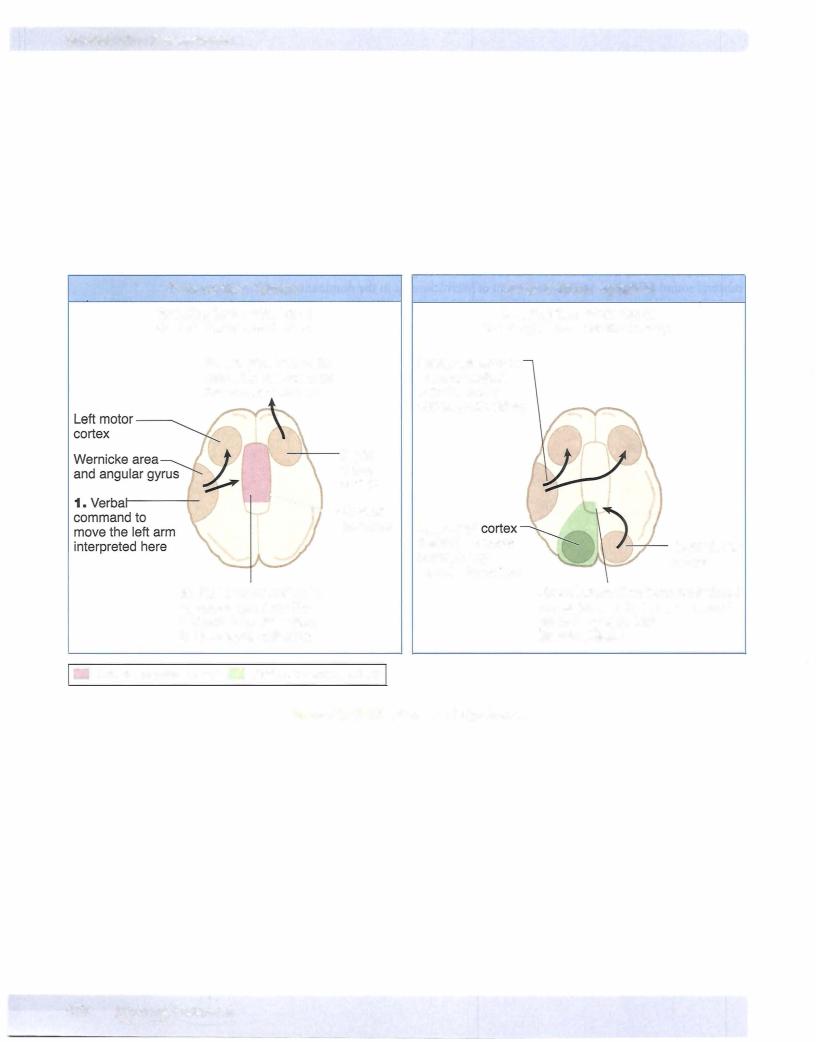
A principal "higher-order" deficit associated with occipital lobe damage is alexia without agraphia (or pure word blindness). The patients are unable to read at all and, curiously, often have a color anomia (inability to name colors). However, they are able to write. This is another example of a disconnect syndrome in which information from the occipital lobe is not available to the parietal or frontal lobes to either understand or express what has been seen.

(Recall that alexia with agraphia-inability to read or write-occurs with lesions encompassing the angular gyrus in the dominant parietal lobe. The cause ofthe syndrome is usually an infarction ofthe left posterior cerebral artery that affects not only the anterior part ofthe occipital lobe but the splenium ofthe corpus callosum. Involvement ofthe left occipital cortex results in a right homonymous hemianopsia with macular sparing. Involvement ofthe splenium ofthe corpus callosum prevents visual information from the intact right occipital cortexfrom reaching language comprehension centers in the left hemisphere. Patients can see words in the left visual field but do not understand what the words mean.
468 MEDICAL

Chapter 10 • Cerebral Cortex

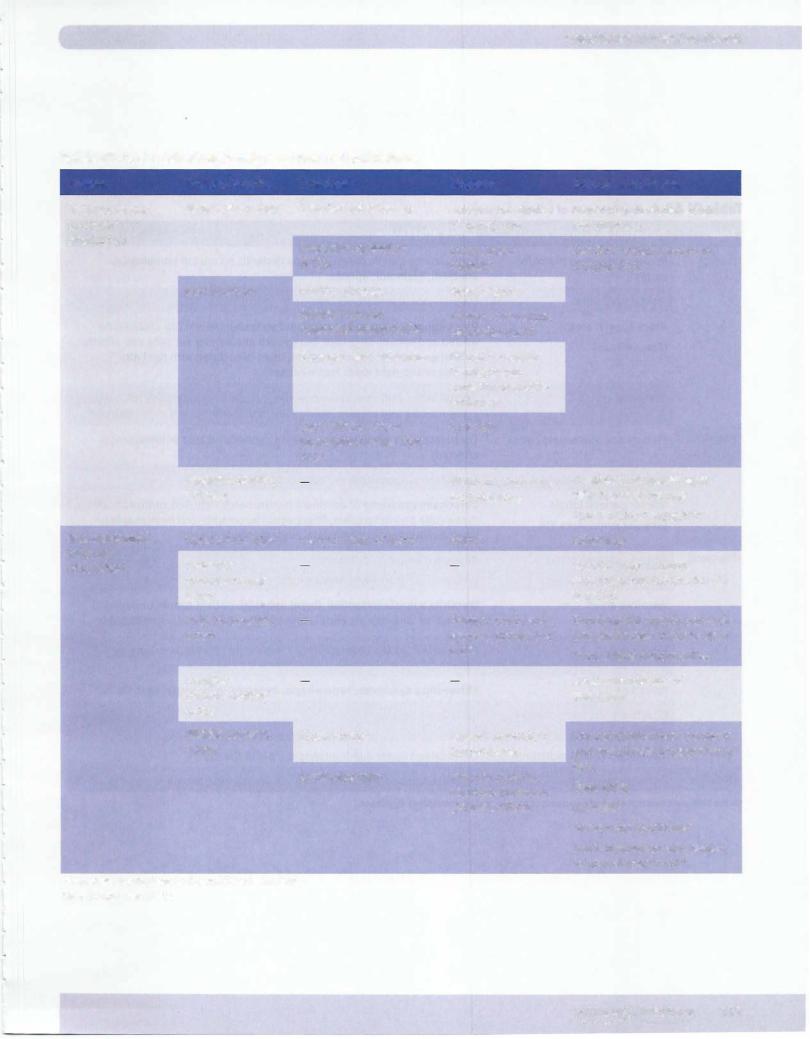
Table IV-10-3. CNS Blood Supply and Stroke-related Deficits

System

Primary Arteries

Branches

Supplies

Deficits after Stroke

Vertebrobasilar

Vertebral arteries

Anterior spinal artery

Anterior two-thirds

Dorsal columns spared; all

(posterior


of spinal cord

else bilateral

circulation)


Posterior cerebellar

Dorsolateral

See Brain-Stem Lesions in


(PICA)

medulla

Chapter IV-5.


Basilar artery

Pontine arteries

Base of pons


Anterior inferior

Inferior cerebellum,


cerebellar artery (AICA)

cerebellar nuclei


Superior cerebellar artery

Dorsal cerebellar


hemispheres;


superior cerebellar


peduncle


Labyrinthine artery

Inner ear


(sometimes arises from


AICA)


Posterior cerebral


Midbrain, thalamus,


arteries


occipital lobe

Internal carotid

Ophthalmic: artery

Central artery of retina

Retina

(anterior

Posterior


circulation)


communicating


artery


Anterior c:erebral


Primary motor and


artery


sensory cortex (leg/


foot)


Anterior


communicating


artery


Middle cerebral

Outer cortical

Lateral convexity of


artery


hemispheres


Lenticulostriate

Internal capsule,


caudate, putamen,


globus pallidus

*If dominant hemisphere is affected (usually the left) tRight parietal lobe lesion

Contralateral hemianopia with macular sparing

Alexia without agraphia*

Blindness

Second most common aneurysm site (often with CN Ill palsy)

Contralateral spastic paralysis and anesthesia of lower limb

Frontal lobe abnormalities

Most common site of aneurysm

Contralateral spastic paralysis and anesthesia of upper limb/ face

Gaze palsy Aphasia*

Gerstmann syndrome""

Hemi inattention and neglect of contralateral bodyt

MEDICAL 471

Источник: https://studfile.net/preview/14638320/