Section IV • Neuroscience
Note
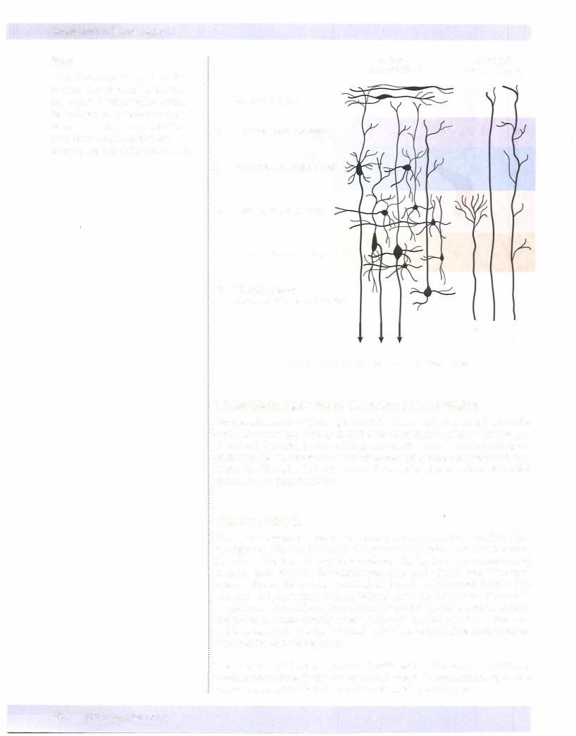
The internal granular layer is the site oftermination of the thalamocortical projections. In primary visual cortex, these fibers form a distinct Line of Gennari. The internal pyramidal layer gives rise to axons that form the corticospinal and corticobulbar tracts.
Efferent |
Afferent |
cortical fibers |
cortical fibers |
I.Molecular layer
II. External granular layer
Ill. External pyramidal layer
IV. Internal granular layer
V. Internal pyramidal layer
VI. Multiform layer
(layer of polymorphic cells)
t t t
Figure IV-1 0-5. The Six-Layered Neocortex
LANGUAGE AND THE DOMINANT HEMISPHERE
Most people (about 80%) are right-handed, which implies that the left side ofthe brain has morehighlydeveloped hand-controlling circuits. In thevast majorityof right-handed people, speech and language functions are also predominantly or ganized in the left hemisphere. Most left-handed people show language functions bilaterally, although a few, with strong left-handed preferences, show right-sided speech and language functions.
BLOOD SUPPLY
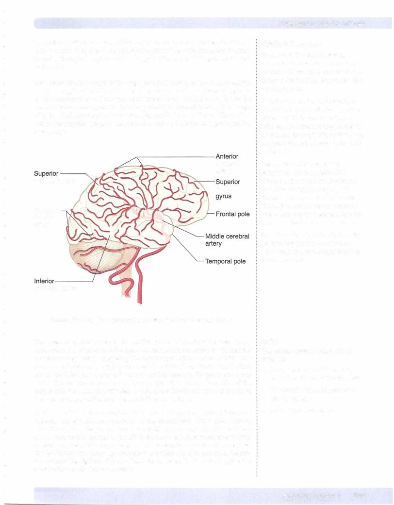
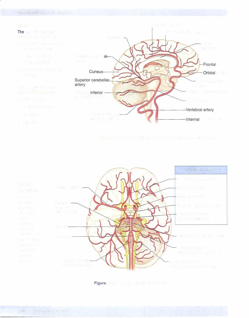
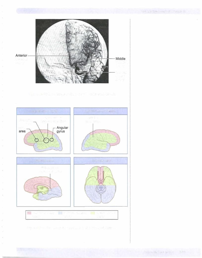
The cortex is supplied by the 2 internal carotid arteries and the 2 vertebral arter ies (Figures IV-10-6 and IV-10-7). On the base (or inferior surface) ofthe brain, branches of the internal carotid arteries and the basilar artery anastomose to form the circle of Willis. The anterior part of the circle lies in front of the optic chiasm, whereas the posterior part is situated just below the mammillary bodies. The circle ofWillis is formedby the terminal part ofthe internal carotid arteries; the proximalparts ofthe anterior and posterior cerebral arteries and the anterior and posterior communicating arteries. The middle, anterior, and posterior cere bralarteries,which arise from the circle ofWillis, supplyall ofthe cerebral cortex, basal ganglia, and diencephalon.
The internal carotid arteryarises from thebifurcation ofthe common carotidand enters the skull through the carotid canal. It enters the subarachnoid space and terminates by dividing into the anterior and middle cerebral arteries.