Section IV • Neuroscience
Clinical Correlate
Receptive Aphasia
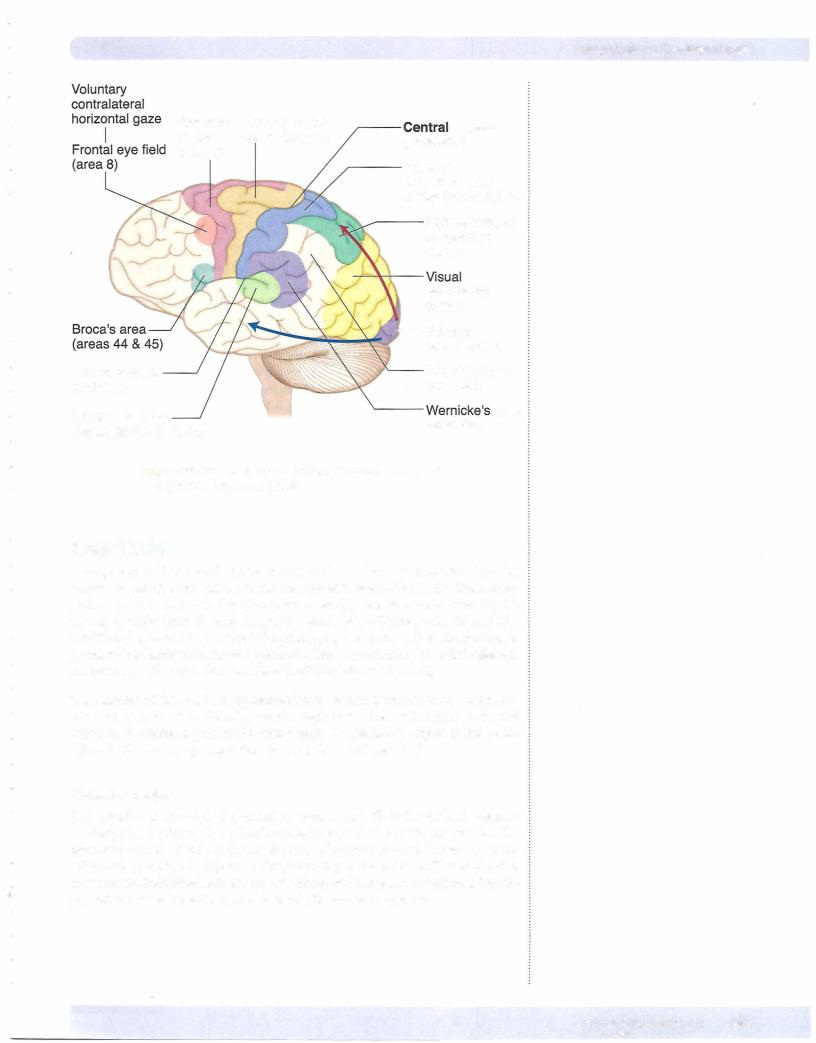
Lesions in area 22 in the temporal lobe and area 39 or 40 in the parietal lobe produce a fluent, receptive, or Wernicke aphasia. The patient with Wernicke aphasia cannot comprehend spoken language and may or may not be able to read (alexia) depending on the extent ofthe lesion. The deficit is characterized by fluent verbalization but lacks meaning. Patients are paraphasic, often misusing words as if speaking using a "word salad."
Patients with Wernicke aphasia are generally unaware oftheir deficit and show no distress as a result oftheir condition.
Gerstmann Syndrome
Ifthe lesion is confined to just the angular gyrus (area 39), the result is a loss of ability to comprehend written language (alexia) and to write it (agraphia), but spoken language may be understood. Alexia with agraphia in pure angular gyrus lesions is often seen with 3 other unique symptoms: acalculia (loss of the ability to perform simple arithmetic problems), finger agnosia (inability to recognize one's fingers), and right-left disorientation. This constellation
of deficits constitutes Gerstmann syndrome and underscores the role of this cortical area in the integration of how children begin to count, add, and subtract using their fingers.
Conduction Aphasia
There is a large fiberbundle connecting areas 22, 39, and 40 with Broca area in the frontal lobe, known as the superior longitudinal fasciculus (or the arcuate fasciculus). A lesion affecting this fiber bundle results in a conduction aphasia. In this patient, verbal output is fluent, but there are many paraphrases and word-finding pauses. Both verbal and visual language comprehension are also normal, but if asked to, the patient cannot repeat words or execute verbal commands by an examiner (such as "Count backwards beginning at 100") and also demonstrates poor object naming. This is an example ofa disconnect syndrome in which the deficit represents an inability to send information from one cortical area to another. As with an expressive aphasia, these patients
are aware ofthe deficit and are frustrated by their inability to execute a verbal command that they fully understand.
Transcortical Apraxia
Lesions to the corpus callosum caused by an infarct ofthe anterior cerebralartery may result in anothertype ofdisconnect syndrome known as a transcortical apraxia. As in other cases ofapraxia, there is no motor weakness, but the patient cannot execute a command to move the left arm. They understand the command, which is perceived in the Wernicke area ofthe left hemisphere, butthe callosal lesion disconnects the Wemicke area from the right primary motor cortex so that the command cannot be executed. The patient is still able to execute a command to move the rightarm because Wernicke area in the left hemisphere is able to communicate with the left primary motor cortexwithout using the corpus callosum.
(Continued)