Материал: 2019 ESC - supraventricular tachycardia
ESC Guidelines |
31 |
Chronic therapy
Catheter ablation is recommended for sympto- |
I |
B |
|
matic, recurrent AVNRT.208,336 339 |
|||
|
|
||
Diltiazem or verapamil, in patients without HFrEF, |
|
|
|
or beta-blockers should be considered if ablation |
IIa |
B |
|
is not desirable or feasible.340 342 |
|
|
|
Abstinence from therapy should be considered |
|
|
|
for minimally symptomatic patients with very |
IIa |
C |
|
infrequent, short-lived episodes of tachycardia.319 |
|
|
i.v. verapamil and diltiazem are contraindicated in the presence of hypotension or HFrEF.
i.v. beta-blockers are contraindicated in the presence of decompensated heart failure.
DC = direct-current; HF = heart failure; HFrEF = heart failure with reduced ejection fraction; i.v. = intravenous.
aClass of recommendation. bLevel of evidence.
|
AVNRT |
|
|
|
|
||
|
Patient out |
|
|
|
|
||
|
of hospital |
|
|
|
|
||
|
Yes |
No |
|
||||
Vagal |
Haemodynamic |
|
|||||
manoeuvres |
instability |
|
|||||
(I B) |
|
|
|
|
|||
|
|
|
No |
|
Yes |
|
|
|
Vagal |
|
Synchronized |
|
|||
|
manoeuvres |
|
cardioversion |
|
|||
|
(I B) |
|
(I B) |
|
|||
|
|
|
|
|
|
|
|
|
If ineffective |
|
|
|
|
|
|
|
|
|
|
|
|
|
|
|
i.v. adenosine |
|
|
|
|
||
|
(I |
|
B) |
|
|
|
|
|
|
|
|
|
|||
|
If ineffective |
|
|
|
|
|
|
|
|
|
|
|
|
|
|
|
|
|
|
|
|||
|
|
|
|
|
|
|
|
i.v. verapamil |
i.v. beta-blocker |
|
|
||||
or diltiazem |
|
|
|||||
(IIa C) |
|
|
|||||
(IIa B) |
|
|
|||||
|
|
|
2019 |
||||
|
|
|
|
|
|
|
|
|
|
|
|
|
|
|
|
|
|
|
|
|
|
|
©ESC |
|
If ineffective |
|
|
|
|||
|
|
|
|
|
|||
|
|
|
|
|
|
|
|
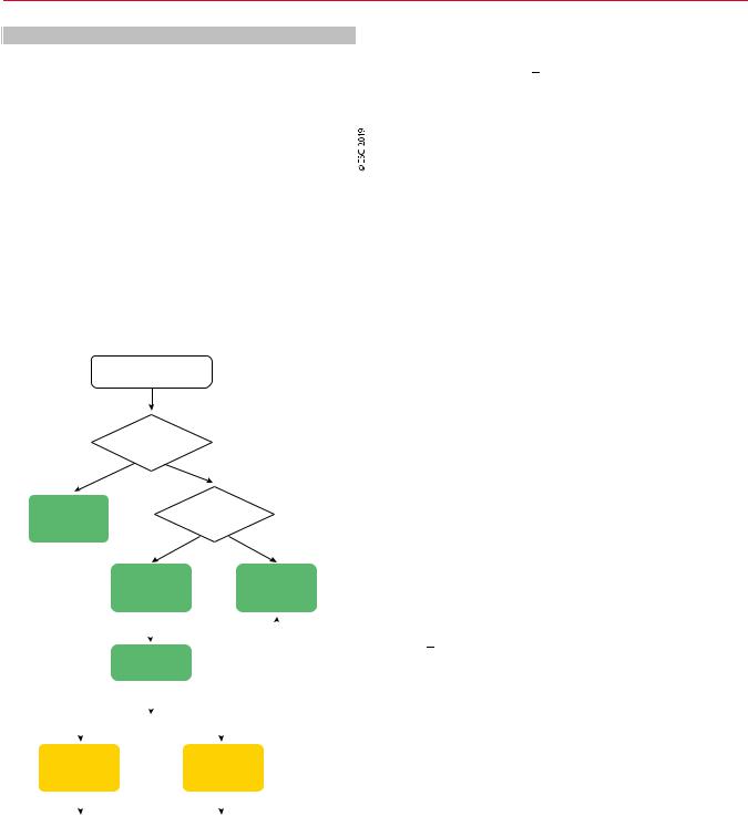
Figure 14 Acute therapy of atrioventricular nodal re-entrant tachycardia.
AVNRT = atrioventricular nodal re-entrant tachycardia; i.v. = intravenous.
11.2.1.2.1 Acute therapy. Most data on the effectiveness of vagal manoeuvres and adenosine for acute termination of tachycardia are derived from mixed populations of SVT, as described in section 10.1.1 on the acute therapy of SVT in general, but it seems that
.
.
.
.
.
.
.
.
.
.
.
.
.
.
.
.
.
.
.
.
.
.
.
.
.
.
.
.
.
.
.
.
.
.
.
.
.
.
.
.
.
.
.
.
.
.
.
.
.
.
.
.
.
.
.
.
.
.
.
.
.
.
.
.
.
.
.
.
.
.
.
.
.
.
.
.
.
.
.
.
.
.
.
.
.
.
.
.
.
.
.
.
.
.
.
.
.
.
.
.
.
.
.
.
.
.
.
.
.
.
.
.
.
.
.
.
.
.
.
.
.
.
.
.
.
.
.
.
.
.
.
.
.
.
.
.
.
.
.
.
.
.
.
.
.
.
.
.
.
.
.
.
.
.
.
.
.
.
.
.
.
.
.
.
.
.
.
.
.
.
.
.
.
they are less successful in AVNRT than in AVRT.89,90,102 A single dose of oral diltiazem (120 mg) plus a beta-blocker (i.e. propranolol 80 mg) may convert <94% of patients, but there is a risk of
hypotension, transient AV block, or—rarely—syncope.342,343 Caution is needed in the elderly, and in patients with known sinus or AV nodal conduction disturbances. A single dose of oral flecainide (3 mg/kg) may also be effective, albeit at a lower rate.342,344 Intranasal etripamil is promising (see section 10.1.1).129 Rarely, when vagal manoeuvres and adenosine cannot terminate the tachycardia and hypotension ensues, synchronized DC cardioversion is indicated101 (Figure 14).
11.2.1.2.2 Catheter ablation. A recent randomized clinical trial (RCT) that compared catheter ablation as first-line treatment with antiarrhythmic drugs demonstrated significant benefits in arrhythmiarelated hospitalizations.336 Furthermore, catheter ablation for SVT in general, and AVNRT in particular, is the current treatment of choice for symptomatic patients because it substantially improves quality of life28,345,346 and reduces costs.347 349 Slow-pathway modification is effective in both typical and atypical AVNRT.338 Usually, a combined anatomical and mapping approach is employed, with ablation lesions delivered at the inferior part of the triangle of Koch, either from the right or the left septal side.337 339,350,351 This approach offers a success rate of 97%, has an 1.3 4% recurrence rate, and has been associated with a risk of AV block of <1% in previous reports.203,204,352,353 Recent experience indicates that in experienced centres, the procedure can be accomplished in both typical and atypical AVNRT with almost no risk of AV block, by targeting the inferior
nodal extension and avoiding the mid-septum, and the roof of the coronary sinus.208,338,354,355 Success rates are lower (82%) and the risk of heart block higher (14%) in patients with adult congenital heart disease (ACHD).356 Usually, recurrences are seen within 3 months following a successful procedure in symptomatic patients who experience frequent episodes of tachycardia,317,329,336,338 but in the young, aged <18 years, recurrences may be seen as long as 5 years
post-ablation.357 IST may occur, but is usually transient and not frequent following slow-pathway ablation.358 Advanced age is not a contraindication for slow-pathway ablation.359 The pre-existence of first-degree heart block carries a higher risk for late AV block and avoidance of extensive slow pathway ablation is preferable
under such conditions.360 There is almost no procedure-related mortality.11,13,203 205,208 Cryoablation may carry a lower risk of AV block, but is associated with a significantly higher recurrence rate.361 363 Its favourable safety profile and higher long-term success rate in younger patients make it especially attractive for the treatment of children.364 AVNRT is a cause of inappropriate shocks in patients with implantable cardioverter defibrillators (ICDs) and, in the case of frequent episodes, catheter ablation is clearly indicated.365
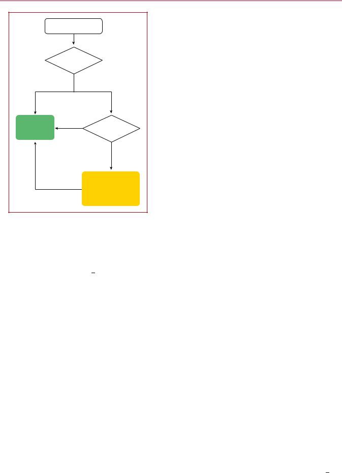
11.2.1.2.3 Chronic therapy. Patients with minimal symptoms and short-lived, infrequent episodes of tachycardia can be followed-up without the need for ablation or long-term pharmacological therapy (Figure 15). Approximately one-half of them may become asymptomatic within the next 13 years.319 Chronic administration of antiarrhythmic drugs decreases the frequency and duration of
2019 September 13 on guest by 1093/eurheartj/ehz467/5556821.abstract/doi/10-article-com/eurheartj/advance.oup.https://academic from Downloaded
32 |
ESC Guidelines |
|
AVNRT |
|
|
|
Symptomatic and |
|
|
|
recurrent |
|
|
|
Yes |
No |
|
Catheter |
No |
Drug therapy |
|
ablation |
|
||
|
desirable |
|
|
(I B) |
|
|
|
|
|
|
|
|
|
Yes |
|
|
|
Diltiazem or verapamil |
|
|
If ineffective |
or beta-blocker |
|
|
|
or diltiazem |
2019 |
|
|
plus beta-blocker |
|
|
|
(IIa B) |
©ESC |
|
|
|
|
Figure 15 Chronic therapy of atrioventricular nodal re-entrant tachycardia.
AVNRT = atrioventricular nodal re-entrant tachycardia.
AVNRT, but has a variable success rate in abolishing tachycardia episodes, ranging from 13 - 82%, and <20% of patients may discon-
tinue therapy.323 In view of the excellent success rate and minimal risk of catheter ablation in symptomatic cases, the value of longterm antiarrhythmic drug therapy seems very limited.
11.2.2 Non-re-entrant junctional tachycardias
Junctional ectopic tachycardia (JET), or focal junctional tachycardia, is an uncommon arrhythmia that arises from abnormal automaticity at the AVN or proximal His bundle. Focal junctional tachycardia in children may be seen as a congenital arrhythmia or, more often, early after infant open-heart surgery.366,367 Congenital junctional tachycardia carries considerable morbidity and mortality.368 Junctional tachycardia can also be seen in adult patients with a structurally normal heart,369,370 and has in the past been associated with non-fibrinolysed acute MI.371 The usual ECG finding in JET is a narrow QRS tachycardia with a short RP interval or AV dissociation. Occasionally, the tachycardia might be irregular and resemble AF.
Propranolol (i.v.) with or without procainamide,370 verapamil, or procainamide,372 or flecainide,373 may be used for acute therapy, but data are scarce. Amiodarone (i.v.) is the drug of choice for post-operative JET as well as for preventing early JET in children after open-heart surgery.374,375 In children with congenital junctional tachycardia, amiodarone alone, or with propafenone or ivabradine, appears effective.368,376 379 For chronic therapy,
.
.
.
.
.
.
.
.
.
.
.
.
.
.
.
.
.
.
.
.
.
.
.
.
.
.
.
.
.
.
.
.
.
.
.
.
.
.
.
.
.
.
.
.
.
.
.
.
.
.
.
.
.
.
.
.
.
.
.
.
.
.
.
.
.
.
.
.
.
.
.
.
.
.
.
.
.
.
.
.
.
.
.
.
.
.
.
.
.
.
.
.
.
.
.
.
.
.
.
.
.
.
.
.
.
.
.
.
.
.
.
.
.
.
.
.
.
.
.
.
.
.
.
.
.
.
.
.
.
.
.
.
.
.
.
.
.
.
.
.
.
.
.
.
.
.
.
.
.
.
.
.
.
.
.
.
.
.
.
.
.
.
.
.
.
.
.
.
.
.
.
.
.
propranolol370 or, in the absence of ischaemic or structural heart disease, flecainide380 and propafenone,381 may be tried. Selective catheter ablation at the site of the earliest retrograde atrial activation is feasible, but carries a lower success rate and higher AV block risk compared with AVNRT (5 - 10%).369,382 Cryoablation is safer.375,383
Non-paroxysmal junctional tachycardia was frequently diagnosed in the past as a junctional rhythm of gradual onset and termination, with a rate between 70 130 b.p.m., and was considered a typical example of digitalis-induced DADs and triggered activity in the AVN.3 The RP interval during tachycardia is variable. Myocardial ischaemia, hypokalaemia, chronic obstructive pulmonary disease, and myocarditis are also associated conditions.
Non-re-entrant AV nodal tachycardia caused by simultaneous multiple nodal pathway conduction (often called double-fire pathology or dual AV nodal tachycardia) is an uncommon mechanism of AV nodal tachycardia,384,385 and has been associated with repetitive retrograde concealment or ‘linking’ phenomena.386 388 These are expressed in the form of ventricular pauses with consistent AV relationship after the pause, and can often be misdiagnosed as AF.389 These extremely rare tachycardias may cause TCM and respond to slow-pathway ablation.
11.3 Atrioventricular arrhythmias
AVRTs use an anatomically defined re-entrant circuit that consists of two limbs: first, the AVN HPS, and second, an AP also called the bypass tract. The two limbs are characterized by differences in refractoriness and conduction times, with critically timed premature atrial or ventricular beats initiating re-entrant tachycardia. On rare occasions, the circuit consists of two APs.
11.3.1 Accessory pathways
APs are single or multiple strands of myocardial cells that bypass the physiological conduction system, and directly connect atrial and ventricular myocardium.390 These AV connections are due to incomplete embryological development of the AV annuli, without complete separation of the atria and ventricles. While there are different types of APs, the most common are those that connect the atrium and the ventricle along the mitral or tricuspid annulus. Approximately 60% are located along the mitral valve and are referred to as left free wall APs, 25% insert along the septal aspect of the mitral or tricuspid annulus, and 15% insert along the right free wall.391 393 Because ventricular muscle is lacking in the proximity of the anterior leaflet of the mitral valve, left-sided APs are usually limited to the region of the mitral annulus at the attachment of the mural (posterior) leaflet. APs located in the superoparaseptal area in close proximity to the His bundle and AVN may also occur.394
APs present characteristic electrophysiological features that differ from AV nodal conduction properties. They typically exhibit fast conduction (with the exception of atypical pathways-see section 11.3.9), dependent on a sodium current similar to that of myocardial cells. Moreover, although a majority of APs conduct both antegradely and retrogradely, some propagate impulses in only one direction. Those that conduct in the antegrade direction only are uncommon (<10%),
2019 September 13 on guest by 1093/eurheartj/ehz467/5556821.abstract/doi/10-article-com/eurheartj/advance.oup.https://academic from Downloaded
ESC Guidelines |
33 |
whereas those that conduct in the retrograde direction only are more frequent (<50%). When the AP conducts antegradely, ventricular pre-excitation is usually evident at rest during sinus rhythm and the AP is referred to as ‘manifest’. Conversely, APs are referred to as ‘concealed’ if they exclusively conduct retrogradely. Concealed APs may have decremental properties.395 The term ‘latent AP’ denotes an AP that is not, or is barely, visible due to location or faster conduction through the AVN.
Multiple APs occur in <12% of patients with pre-excitation, and in
<50% in patients with Ebstein’s anomaly.396
AVRT is the most common tachycardia associated with APs. Two mechanisms of re-entry are possible according to the antegrade or retrograde conduction over the AVN HPS, and are classified as orthodromic and antidromic AVRT.
11.3.2 Wolff Parkinson White syndrome
WPW syndrome refers to the presence of an overt (manifest) AP, thus resulting in the so-called pre-excitation, in combination with usually recurrent tachyarrhythmias.397 During sinus rhythm, a typical pattern in the resting ECG with the following characteristics is present: (i) a short PR interval (<120 ms); (ii) slurred upstroke (or downstroke) of the QRS complex (‘delta wave’); and (iii) a wide
.
.
.
.
.
.
.
.
.
.
.
.
.
.
.
.
.
.
.
.
.
.
.
.
.
.
.
.
.
.
.
.
.
.
.
.
.
.
.
.
.
.
.
.
.
.
.
.
.
.
.
.
.
.
.
.
.
.
.
.
.
.
.
.
.
.
.
QRS complex (>120 ms). In most cases, APs giving rise to the WPW pattern are seen in structurally normal hearts. Rare, familial forms of pre-excitation associated with LV hypertrophy and multisystem disease [mutations in the protein kinase adenosine monophosphate-activated non-catalytic subunit gamma 2 (PRKAG2) gene, Danon and Fabry disease, and others] have also been described.398
Several surface ECG algorithms have been developed that can be
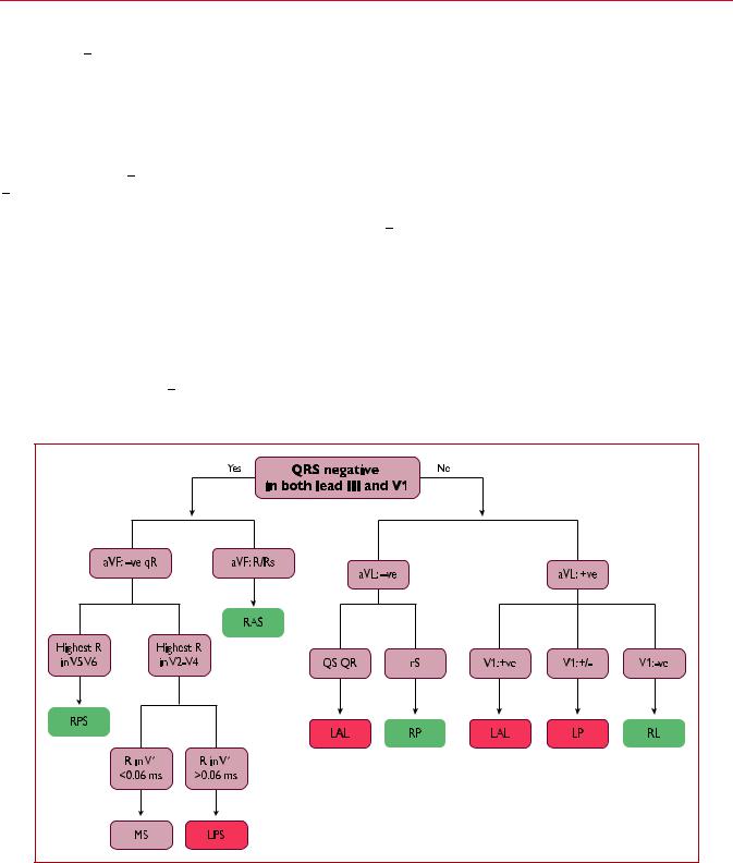
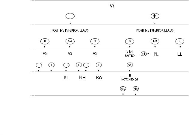
applied for the localization of APs in the presence of overt preexcitation (Figures 16 and 17).399 401 Pre-excitation on the surface ECG can be intermittent and can even disappear permanently (in <35% of cases) over time. Furthermore, various degrees of preexcitation are possible depending on the location of the AP as well as on AVN conduction properties.
11.3.3 Orthodromic atrioventricular re-entrant tachycardia
Orthodromic AVRT accounts for >90% of AVRTs and for 20 - 30% of all sustained SVTs. The re-entrant impulse conducts from the atrium to the ventricle through the AVN HPS, which is the anterograde limb of the re-entrant circuit, whereas the AP conducts from the ventricle to the atrium, and serves as the
Figure 16 The St George’s algorithm for the localization of accessory pathways.399 þve = QRS complex-positive; ve = QRS complex-negative; þ/- = QRS complex equiphasic; AP = accessory pathway; LAL = left anterolateral; LP = left posterior; LPL = left posterolateral; LPS = left posteroseptal; MS = mid-septal; RAS = right anteroseptal; RL = right lateral; RP = right posterior; RPS = right posteroseptal.
2019 September 13 on guest by 1093/eurheartj/ehz467/5556821.abstract/doi/10-article-com/eurheartj/advance.oup.https://academic from Downloaded
34 |
|
|
|
|
|
|
|
|
|
|
|
|
|
|
|
|
|
|
|
|
|
|
|
|
|
|
|
|
|
|
|
|
|
|
|
|
|
|
|
|
|
|
|
|
|
|
|
ESC Guidelines |
||
|
|
|
|
|
|
|
|
|
|
|
|
|
|
|
|
|
|
|
|
|
|
|
|
|
|
|
|
|
|
|
|
|
|
|
|
|
|
|
|
|
|
|
|
|
|
|
|
|
|
|
|
|
|
|
|
|
|
|
|
|
|
|
|
|
|
|
|
|
|
|
|
|
|
|
|
|
|
|
|
|
|
|
|
|
|
|
|
|
|
|
|
|
|
|
|
|
|
|
|
|
|
|
|
|
|
|
|
|
|
|
|
|
|
|
|
|
|
|
|
|
|
|
|
|
|
|
|
|
|
|
|
|
|
|
|
|
|
|
|
|
|
|
|
|
|
|
|
|
|
|
|
|
|
|
|
|
|
|
|
|
|
|
|
|
|
|
|
|
|
|
|
|
|
|
|
|
|
|
|
|
|
|
|
|
|
|
|
|
|
|
|
|
|
|
|
|
|
|
|
|
|
|
|
|
|
|
|
|
|
|
|
|
|
|
|
|
|
|
|
|
|
|
|
|
|
|
|
|
|
|
|
|
|
|
|
|
|
|
|
|
|
|
|
|
|
|
|
|
|
|
|
|
|
|
|
|
|
|
|
|
|
|
|
|
|
|
|
|
|
|
|
|
|
|
|
|
|
|
|
|
|
|
|
|
|
|
|
|
|
|
|
|
|
|
|
|
|
|
|
|
|
|
|
|
|
|
|
|
|
|
|
|
|
|
|
|
|
|
|
|
|
|
|
|
|
|
|
|
|
|
|
|
|
|
|
|
|
|
|
|
|
|
|
|
|
|
|
|
|
|
|
|
|
|
|
|
|
|
|
|
|
|
|
|
|
|
|
|
|
|
|
|
|
|
|
|
|
|
|
|
|
|
|
|
|
|
|
|
|
|
|
|
|
|
|
|
|
|
|
|
|
|
|
|
|
|
|
|
|
|
|
|
|
|
|
|
|
|
|
|
|
|
|
|
|
|
|
|
|
|
|
|
|
|
|
|
|
|
|
|
|
|
|
|
|
|
|
|
|
|
|
|
|
|
|
|
|
|
|
|
|
|
|
|
|
|
|
|
|
|
|
|
|
|
|
|
|
|
|
|
|
|
|
|
|
|
|
|
|
|
|
|
|
|
|
|
|
|
|
|
|
|
|
|
|
|
|
|
|
|
|
|
|
|
|
|
|
|
|
|
|
|
|
|
|
|
|
|
|
|
|
|
|
|
|
|
|
|
|
|
|
|
|
|
|
|
|
|
|
|
|
|
|
|
|
|
|
|
|
|
|
|
|
|
|
|
|
|
|
|
|
|
|
|
|
|
|
|
|
|
|
|
|
|
|
|
|
|
|
|
|
|
|
|
|
|
|
|
|
|
|
|
|
|
|
|
|
|
|
|
|
|
|
|
|
|
|
|
|
|
|
|
|
|
|
|
|
|
|
|
|
|
|
|
|
|
|
|
|
|
|
|
|
|
|
|
|
|
|
|
|
|
|
|
|
|
|
|
|
|
|
|
|
|
|
|
|
|
|
|
|
|
|
|
|
|
|
|
|
|
|
|
|
|
|
|
|
|
|
|
|
|
|
|
|
|
|
|
|
|
|
|
|
|
|
|
|
|
|
|
|
|
|
|
|
|
|
|
|
|
|
|
|
|
|
|
|
|
|
|
|
|
|
|
|
|
|
|
|
|
|
|
|
|
|
|
|
|
|
|
|
|
|
|
|
|
|
|
|
|
|
|
|
|
|
|
|
|
|
|
|
|
|
|
|
|
|
|
|
|
|
|
|
|
|
|
|
|
|
|
|
|
|
|
|
|
|
|
|
|
|
|
|
|
|
|
|
|
|
|
|
|
|
|
|
|
|
|
|
|
|
|
|
|
|
|
|
|
|
|
|
|
|
|
|
|
|
|
|
|
|
|
|
|
|
|
|
|
|
|
|
|
|
|
|
|
|
|
|
|
|
|
|
|
|
|
|
|
|
|
|
|
|
|
|
|
|
|
|
|
|
|
|
|
|
|
|
|
|
|
|
|
|
|
|
|
|
|
|
|
|
|
|
|
|
|
|
|
|
|
|
|
|
|
|
|
|
|
|
|
|
|
|
|
|
|
|
|
|
|
|
|
|
|
|
|
|
|
|
|
|
|
|
|
|
|
|
|
|
|
|
|
|
|
|
|
|
|
|
|
|
|
|
|
|
|
|
|
|
|
|
|
|
|
|
|
|
|
|
|
|
|
|
|
|
|
|
|
|
|
|
|
|
|
|
|
|
|
|
|
|
|
|
|
|
|
|
|
|
|
|
|
|
|
|
|
|
|
|
|
|
|
|
|
|
|
|
|
|
|
|
|
|
|
|
|
|
|
|
|
|
|
|
|
|
|
|
|
|
|
|
|
|
|
|
|
|
|
|
|
|
|
|
|
|
|
|
|
|
|
|
|
|
|
|
|
|
|
|
|
|
|
|
|
|
|
|
|
|
|
|
|
|
|
|
|
|
|
|
|
|
|
|
|
|
|
|
|
|
|
|
|
|
|
|
|
|
|
|
|
|
Figure 17 Localization of accessory pathways in the presence of maximum (spontaneous or evoked) pre-excitation.400 Accessory pathway locations are green when right-sided and red when left-sided. Left posterolateral accessory pathways can have 0, 1, or 2 inferior leads with positive polarity, whereas nodo-Hisian accessory pathways can have 1, 2, or 3 inferior leads with positive polarity. Right-sided accessory pathways are framed orange or yellow when the V3 lead is negative or positive, respectively. Left posterior accessory pathways are framed blue when the V1/I ratio is <1 or purple when V1/I ratio is >1. AP = accessory pathway; DCS = deep coronary sinus; LL = left lateral; LPL = left posterolateral; LPS = left paraseptal; NH = nodo-Hisian; RA = right anterior; RL = right lateral; RP = right posterior; RPS = right paraseptal.
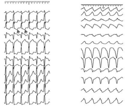
retrograde limb of the re-entrant circuit. Orthodromic AVRT tends to be a rapid tachycardia, with frequencies ranging from 150 to, rarely, >220 b.p.m. During tachycardia (Figure 18), the following ECG features can be present: (i) RP interval constant and, usually but not invariably, up to one-half of the tachycardia CL; (ii) narrow QRS; (iii) functional BBB usually associated with an AP ipsilateral to the blocked bundle, especially in young patients (aged <40 years); and (iv) ST-segment depression.
11.3.4 Antidromic atrioventricular re-entrant tachycardia
Antidromic AVRT occurs in 3 - 8% of patients with WPW syndrome.402 404 The re-entrant impulse travels from the atrium to the ventricle through the AP with anterograde conduction; meanwhile, retrograde conduction occurs over the AVN or another AP, usually located in a contralateral position to ensure longer travel distances, thus allowing for sufficient recovery of refractoriness of the respective elements of the re-entrant circuit. In 30 - 60% of patients with spontaneous antidromic AVRT, multiple APs (manifest or concealed), which could act or not as the retrograde limb during the AVRT, may be detected. Antidromic AVRT has the following ECG features, illustrated in Figure 18: (i) a wide QRS complex (fully pre-excited) and (ii) an RP interval that is
.
.
.
.
.
.
.
.
.
.
.
.
.
.
.
.
.
.
.
.
.
.
.
.
.
.
.
.
.
.
.
.
.
.
.
.
.
.
.
.
.
.
.
.
.
.
.
.
.
.
.
.
.
.
.
.
.
.
.
.
.
.
.
.
.
.
.
.
.
.
.
.
difficult to assess as the retrograde P wave is usually inscribed within the ST-T segment.
11.3.5 Accessory pathway as a bystander
In the presence of focal AT, atrial flutter, AF, or AVNRT, the QRS complexes can be pre-excited when the AP acts as a bystander, and is not a critical part of the re-entry circuit.
11.3.6 Pre-excited atrial fibrillation
Paroxysmal AF has been found in 50% of patients with WPW, and may be the presenting arrhythmia in affected patients.405,406 These patients are typically young and have no structural heart disease. High-rate AVRT may potentially initiate AF. AF with fast ventricular response over an overt AP with a short anterograde refractory period is a potentially life-threatening arrhythmia in patients with WPW syndrome, due to potential degeneration into VF.
11.3.7 Concealed accessory pathways
Concealed APs give rise only to orthodromic AVRT. Their true prevalence is unknown because they are not detectable on the resting surface ECG, but only at occurrence of AVRT, or during electrophysiology testing.45 No sex predilection is found and these pathways
2019 September 13 on guest by 1093/eurheartj/ehz467/5556821.abstract/doi/10-article-com/eurheartj/advance.oup.https://academic from Downloaded
ESC Guidelines |
35 |
|
|
I |
I |
|
II |
|
|
|
|
|
II |
III |
|
|
|
|
III |
aVR |
|
|
|
|
aVR |
aVL |
|
aVL |
aVF |
|
aVF |
V1 |
|
|
|
|
V1 |
V2 |
|
|
|
|
V2 |
V3 |
|
|
|
|
V3 |
|
|
|
V4 |
|
V4 |
|
|
V5 |
V5 |
|
|
2019 |
|
V6 |
V6 |
|
|
©ESC |
|
|
|
Figure 18 Atrioventricular re-entrant tachycardia. Left: othodromic atrioventricular re-entrant tachycardia due to a concealed posteroseptal accessory pathway. Retrograde P waves are negative during tachycardia in the inferior leads (arrows). Right: Antidromic atrioventricular re-entrant tachycardia due to an atriofascicular accessory pathway. The axis during tachycardia due to atypical pathways depends on the way of insertion into the right bundle and fusion over the left anterior fascicle.
tend to occur more frequently in younger patients than in those with AVNRT; however, significant overlap exists.3 Concealed APs are predominantly localized along the left free wall (64%), and less frequently at septal (31%) and right free wall locations.395 Clinical presentation is with AVRT. Concealed pathways are not associated with an increased risk of sudden cardiac death. The management of AVRT due to a concealed AP is similar to that of an overt AP, but in this case is related to symptoms without significant prognostic relevance in most cases.
11.3.8 Permanent junctional reciprocating tachycardia
PJRT is a rare form of AV reciprocating tachycardia using a concealed AP. Usually these APs, originally described by Coumel, are located in the posteroseptal region and are associated with retrograde decremental conduction properties.407 PJRT is a long RP tachycardia due to the slow conduction properties of the AP, and is characterized by deeply inverted retrograde P waves in leads II, III, and aVF due to the retrograde nature of atrial activation. The incessant nature of PJRT may result in TCM that usually resolves after successful treatment by radiofrequency catheter ablation, particularly in younger patients.407,408 Catheter ablation is strongly recommended in symptomatic patients or in cases with impaired LV ejection fraction likely related to TCM.
.
.
.
.
.
.
.
.
.
.
.
.
.
.
.
.
.
.
.
.
.
.
.
.
.
.
.
.
.
.
.
.
.
.
.
.
.
.
.
.
.
.
.
.
.
.
.
.
.
.
.
.
.
.
.
.
.
.
.
.
.
.
.
.
.
.
.
.
.
.
.
.
.
Other potential causes of long RP tachycardias are sinus tachycardia, AT, atypical AVRT, and JET with 1:1 retrograde conduction.
11.3.9 Atypical forms of pre-excitation
Other APs are postulated to result in cardiac pre-excitation. Atypical APs (also called Mahaim fibers) are connections between the right atrium or the AVN and the right ventricle, into or close to the right bundle branch.409 414 Most of them are atriofascicular or nodoventricular (as initially described), but they can also be atriofascicular, atrioventricular, nodofascicular, or nodoventricular, depending on their variable proximal and distal insertions.413,414 Left-sided atypical pathways have also been described but are extremely rare.415 417
Atypical pathways usually contain accessory nodal tissue, which results in decremental properties, and connect the atrium to the fascicles by crossing the lateral aspect of the tricuspid annulus, but posteroseptal locations can also be found in rare cases. Conduction is usually anterograde only, but concealed fibres have also been described.412,418 The following properties define the behaviour of atypical pathways:
•Baseline normal QRS or different degrees of manifest preexcitation with LBBB morphology;
•Programmed atrial pacing, leading to obvious manifest preexcitation following an increase in AV interval along with shortening of the HV interval at shorter pacing CLs;
2019 September 13 on guest by 1093/eurheartj/ehz467/5556821.abstract/doi/10-article-com/eurheartj/advance.oup.https://academic from Downloaded