Материал: 2019 ESC - supraventricular tachycardia
26 |
ESC Guidelines |
|
|
Atrial flutter/MRAT
|
|
Haemodynamic |
|
|
||
|
|
|
instability |
|
|
|
|
No |
|
|
|
Yes |
|
|
|
|
|
|
Synchronized |
|
|
Rhythm control |
|
|
|
cardioversion |
|
|
|
|
|
|
(I B) |
|
|
strategy |
|
|
|
|
|
|
|
|
|
|
|
|
No |
Yes |
|
|
|
||
i.v. beta-blocker |
Electrical |
|
|
|
||
or |
|
|
|
|||
i.v. diltiazem or verapamil |
cardioversion |
|
|
|
||
(IIa B) |
preferred |
|
|
|
||
|
Yes |
No |
|
|
||
Low-energy |
|
|
|
PPM/ICD |
||
synchronized cardioversion |
|
|
|
|||
|
(I B) |
|
|
|
present? |
|
|
|
|
|
No |
|
Yes |
|
|
i.v. ibutilide |
|
|
Ηigh-rate |
|
|
or dofetilide i.v. or |
|
|
|||
|
|
|
atrial pacing |
|||
|
oral (in-hospital) |
|
|
|||
|
|
|
(I B) |
|||
|
|
(I B) |
|
|
||
|
|
|
|
|
||
|
If not available |
|
|
|
|
|
|
or contra-indicated |
|
|
|
|
|
|
|
|
|
|
|
|
|
|
|
|
|
|
|
i.v. amiodarone |
Invasive or non-invasive |
2019 |
|
high-rate atrial pacing |
|||
(IIb C) |
|||
|
|
||
|
(IIb B) |
©ESC |
|
|
|
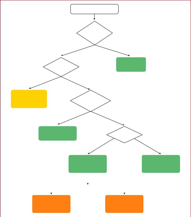
Figure 11 Acute therapy of stable atrial flutter or macro-re-entrant atrial tachycardia.
ICD = implantable cardioverter defibrillator; i.v. = intravenous; MRAT = macro-re-entrant atrial tachycardia; PPM: permanent pacemaker.
2019 September 13 on guest by 1093/eurheartj/ehz467/5556821.abstract/doi/10-article-com/eurheartj/advance.oup.https://academic from Downloaded

ESC Guidelines |
27 |
flutter and even the combination of AVN-blocking drugs (digoxin, beta-blockers, and calcium channel blockers) may fail, making cardioversion to sinus rhythm necessary. Dofetilide and ibutilide, pure class III antiarrhythmic drugs, are generally effective in interrupting atrial flutter in i.v. administration (dofetilide may be also given orally for this purpose), while class IA and IC drugs have little or no effect. Class IC antiarrhythmic drugs should not be used in the absence of AV-blocking agents because of the risk of slowing the atrial rate, which may result in 1:1 AV conduction. Amiodarone may not be very effective acutely to reestablish sinus rhythm, but it does help to control the ventricular rate if it is too fast.275,276 Low-energy electrical cardioversion is commonly used with haemodynamic compromise or after failure of previous actions, but it could be the first choice due to its high efficacy. Electrical cardioversion for atrial flutter is more effective and less energy is required, compared with AF. When atrial electrodes are in place, high-rate stimulation can be used to convert flutter, sometimes through AF.258,259 If pacing induces AF, this may allow better control of the ventricular rate than flutter. Atrial stimulation can also be done with percutaneous endocardial electrodes or from the oesophagus; this is mostly done in paediatrics.261 Pre-treatment with procainamide may facilitate conversion of atrial flutter by atrial pacing.277 Data on pre-cardioversion anticoagulation are lacking, but most probably patients should be treated the same as those with AF.4,278
11.1.4.1.4 Catheter ablation. Catheter ablation is the most effective therapy to maintain sinus rhythm, and is clearly superior to amiodarone.262,263 Ablation of CTI with confirmed bidirectional conduction block results in a <10% rate of recurrence.279 However, the incidence of AF is high in the long-term.280 When typical CTIdependent atrial flutter ensues during antiarrhythmic drug therapy (class IC or amiodarone) for AF, CTI ablation is a reasonable
choice to ensure that antiarrhythmic drugs can be continued for AF control.262,263
Although no procedure-related mortality had been detected in early studies,203,204 in recent studies, mortality and stroke rates of 0.2 - 0.34 and 0.19 - 0.5%, respectively, have been reported (Table 11).12,206 In a recent registry, ablation for flutter had a higher mortality than that for AF (0.3 vs. 0.05%), but this might have been due to the comorbidities or advanced ages of patients referred for flutter ablation.207
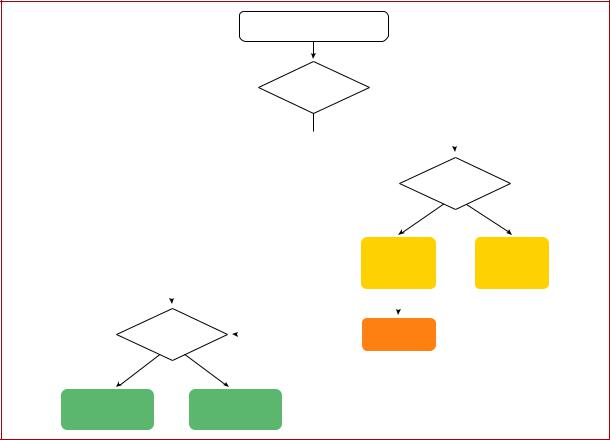
11.1.4.1.5Chronic therapy. Rate control is part of the therapeutic approach, using AV nodal blocking agents such as diltiazem, verapamil, or beta-blockers (Figure 12). When ablation is not feasible or the
patient’s preference, antiarrhythmic drugs may also be used to maintain sinus rhythm. Dofetilide257 and sotalol281 are useful, but there are concerns about pro-arrhythmia. Amiodarone may have a role,263 but it
should be restricted to cases of HF or significant structural heart disease.
11.1.4.1.6Anticoagulation. Data about the embolic risk of atrial flutter have usually been derived in the presence of concomitant AF, thus making individualized risk stratification difficult. Left atrial (LA) appendage ‘stunning’ and thrombi seem to be lower
.
.
.
.
.
.
.
.
.
.
.
.
.
.
.
.
.
.
.
.
.
.
.
.
.
.
.
.
.
.
.
.
.
.
.
.
.
.
.
.
.
.
.
.
.
.
.
.
.
.
.
.
.
.
.
.
.
.
.
.
.
.
.
.
.
.
.
.
.
.
.
.
.
.
.
.
.
.
.
.
.
.
.
.
.
.
.
.
.
.
.
.
.
.
.
.
.
.
.
.
.
.
.
.
.
.
.
.
.
.
.
.
.
.
.
.
.
.
.
.
.
.
.
.
.
.
.
.
.
.
.
.
.
.
.
.
.
.
.
.
.
.
.
.
.
.
.
.
.
.
.
.
.
.
.
.
.
.
.
.
.
.
.
.
.
.
.
.
compared with those in AF.247,282 The thrombo-embolic risk of atrial flutter, although lower than that of AF,246 is still significant.241 244 That, together with the association with AF, justifies thromboprophylaxis, and anticoagulation has been recommended as in AF.2,3 These recommendations extend to the acute setting for cardioversion when flutter lasts for >48 h.278 However, it should be noted that there is a lack of prospective, dedicated, randomized studies on the subject. Furthermore, the value of the CHA2DS2-VASc [Cardiac failure, Hypertension, Age >75 (Doubled), Diabetes, Stroke (Doubled) Vascular disease, Age 65 74 and Sex category (Female)] score in preventing ischaemic stroke in patients with atrial flutter has not been established,245 and in patients without concomitant AF the threshold for the initiation of anticoagulation appears to be higher than that for patients with AF.246
11.1.4.1.7 Other cavotricuspid isthmus-dependent macro re-
An atypical ECG pattern may not exclude CTI-dependent MRAT.283 Lower-loop re-entry refers to a circuit rotating around the inferior vena cava instead of around the tricuspid annulus. It may be clockwise or counter-clockwise.284,285 When rotating counter-clockwise, it might be considered a variant of typical counter-clockwise flutter with a caudal shift of the cranial turning point posterior to the entry of the superior vena cava, resulting in a similar ECG appearance. ‘Figure-of-eight double-loop reentry’ may also occur around the inferior vena cava and tricuspid annulus, and mimic typical clockwise atrial flutter.285 Other circuits using part of the CTI or even restricted inside it, are in essence CTI-
dependent with a similar ECG appearance to typical common flutter.286,287
11.1.4.2 Non-cavotricuspid isthmus-dependent macro re-entrant atrial tachycardia
The terms non-CTI-dependent MRAT and atypical flutter are used interchangeably, and describe flutter waves in the ECG not suggestive of typical circuits. The pitfall with this use comes from the atypical ECG that may happen when typical circuits develop in diseased atria, most frequently after surgery or extensive ablation, or under the effects of antiarrhythmic drugs. Conversely, upperloop re-entry may mimic a typical flutter ECG pattern without being CTI-dependent.283 True atypical flutter is actually a post hoc diagnosis when the circuit has been outlined and dependence on CTI has been ruled out.
11.1.4.2.1 Right atrium macro2re-entrant atrial tachycardia. Atrial sutures and patches used for complex congenital heart disease surgery, together with progressive atrial damage, create multiple obstacles and protected isthmuses that constitute the substrate for complex and multiple MRAT.288,289 This usually happens around RA free wall scars. However, in patients with complex congenital heart disease, the presence of extensive atrial scars hinders the differential diagnosis of focal or MRAT.290
Figure-of-eight double-loop tachycardias mimicking the ECG pattern of a common atrial flutter may also occur following surgical atriotomy.291
2019 September 13 on guest by 1093/eurheartj/ehz467/5556821.abstract/doi/10-article-com/eurheartj/advance.oup.https://academic from Downloaded
28 |
ESC Guidelines |
|
|
Atrial flutter/MRAT
Symptomatic and
recurrent
Yes |
No |
|
|
|
|
|
|
|
|
|
|
|
|
|
|
|
|
Drug therapy |
|
|
|
|
|
|
|
|
desirable |
|
|
|
|
|
|
Yes |
|
No |
|
|
|
|
|
|
Beta-blocker |
|
Catheter |
|
|
|
|
|
|
or diltiazem |
|
||
|
|
|
|
|
|
ablation |
||
|
|
|
|
|
or verapamil |
|
||
|
|
|
|
|
|
(IIa B) |
||
|
|
|
|
|
(IIa C) |
|
||
|
|
|
|
|
|
|
||
|
|
|
|
If ineffective |
|
|
|
|
|
|
|
|
|
|
|||
|
|
|
|
|
|
|
|
|
CTI dependent |
|
|
|
Amiodarone |
|
|
||
flutter |
|
If ineffective |
|
(IIb C) |
|
|
||
|
|
|
|
|
|
|
|
|
Yes |
|
or not tolerated |
|
|
|
|
|
|
No |
|
|
|
|
|
|||
Catheter |
Catheter ablation |
|
|
|
2019 |
|||
|
|
|
|
|
||||
ablation |
in experienced |
|
|
|
|
©ESC |
||
(I A) |
centres (I B) |
|
|
|
|
|||
|
|
|
|
|
||||
Figure 12 Chronic therapy of atrial flutter/macro-re-entrant atrial tachycardia.
CTI = cavotricuspid isthmus; MRAT = macro-re-entrant atrial tachycardia.
RA MRAT may also occur in the absence of previous interven-
tion. Most of these are sustained around areas of ‘electrical silence’ in the RA free wall, probably due to fibrosis.224,264,266 Atypical atrial flutter could also arise from upper loop re-entry in the right atrium with conduction through the gap in the crista terminalis.269
Rate control is often difficult due to the regularity and usually slow rate of the tachycardia. Antiarrhythmic drugs are often ineffective, or their use is limited because of structural heart disease and comorbidities. Radiofrequency ablation of often several critical isthmuses is the most effective treatment. Circuits around scars of longitudinal atriotomy can be mapped and ablated with good long-term results.267,292 However, owing to the complexity of possible substrates and difficulty of reaching critical isthmuses, ablation procedures for these patients should be restricted to experienced operators and centres.
11.1.4.2.2 Left atrium macro2re-entrant atrial tachycardia. Circuits sustaining LA atypical flutter/MRAT are most usually due to electrically silent areas of abnormal tissue, following medical interventions or progressive atrial degeneration/fibrosis.268 Anatomical obstacles such as the ostia of PVs, and mitral annulus, are often involved.
.
.
.
.
.
.
.
.
.
.
.
.
.
.
.
.
.
.
.
.
.
.
.
.
.
.
.
.
.
.
.
.
.
.
.
.
.
.
.
.
.
.
.
.
.
.
.
.
.
.
.
.
.
.
.
.
.
.
.
.
.
.
.
.
.
.
.
.
.
Owing to its widespread use, AF ablation is the procedure that frequently causes the lesions able to sustain re-entry circuits, usually after linear ablation or extensive defragmentation. Pre-existing atrial disease is also predictive of macro-re-entry.293 Localized segmental PV dis-
connection may cause focal tachycardias,294 and circumferential antral ablation may also create MRAT due to gaps in the lines.295 299 AT due to a small re-entrant circuit after ablation of AF may possibly be distinguished from macro-re-entry by a shorter P-wave duration. RA MRATs have a higher incidence of negative polarity in at least one precordial lead compared with LA macro-re-entry.300 302
Atrial circuits are also created after surgery for different conditions, including mitral valve disease, and are related to incisions or cannulation.303 Surgery to treat AF may also result in macro-re-entry circuits and focal AT.304
Circuits causing atypical left MRAT may also occur in the LA without prior intervention, commonly, but not invariably, associated with significant left heart disease.305 These are based on areas of electrical silence, probably due to fibrosis, engaging anatomical obstacles such as the ostia of the PVs or the mitral annulus, and may be ablated by interrupting critical isthmuses.265,306 Circuits may also happen in the LA septum due to slow conduction caused by atrial disease or antiarrhythmic drugs.307
2019 September 13 on guest by 1093/eurheartj/ehz467/5556821.abstract/doi/10-article-com/eurheartj/advance.oup.https://academic from Downloaded
ESC Guidelines |
29 |
Peri-mitral flutter, sometimes incorporating silent areas at the roof of the LA, is ablated in a similar way to peri-tricuspid circuits. However, the deployment of a stable line of block at critical isthmuses is more challenging.308 310 Circuits around the PVs are also frequently recognized and ablated.280,295,296 Intervention to treat these tachycardias after the initial procedure should be delayed, if possible, for >3 months. As part of the maturation process of the deployed lesions, some tachycardias may be transient in nature,311 and initial rate control and/or use of antiarrhythmic drugs is favoured.
11.2 Atrioventricular junctional arrhythmias
11.2.1 Atrioventricular nodal re-entrant tachycardia
AVNRT denotes re-entry in the area of the AVN, but the exact circuit remains elusive. The AVN is a three-dimensional structure with greater variability in the space constant of tissue, and poor gap junction connectivity due to differential expression of connexin isoforms, conditions that provide an explanation for dual conduction and nodal re-entrant arrhythmogenesis.312 314 There has also been considerable histological and electrophysiological evidence that the right and left inferior extensions of the human AVN, and the atrionodal inputs that they facilitate, may provide the anatomical substrate for the slow pathway.315,316 Thus, comprehensive models of the tachycardia
.
.
.
.
.
.
.
.
.
.
.
.
.
.
.
.
.
.
.
.
.
.
.
.
.
.
.
.
.
.
.
.
.
.
.
.
.
.
.
.
.
.
.
.
.
.
.
.
.
.
.
.
.
.
.
.
.
.
.
.
.
.
.
.
.
.
.
.
.
.
.
.
.
.
circuit for all forms of AVNRT based on the concept of atrionodal inputs have been proposed.47,317
Onset of AVNRT seems to occur bimodally over time. In many patients, attacks indeed manifest early in life, whereas in a substantial proportion of patients AVNRT starts later, e.g. in the fourth or fifth decade of life.318 One-half of the patients with minimal symptoms and short-lived, infrequent episodes of tachycardia may become asymptomatic within the next 13 years.319 AVNRT may result in AF that usually, although not invariably, is eliminated following catheter ablation of AVNRT.320 Familial AVNRT should be considered.321
11.2.1.1 Diagnosis
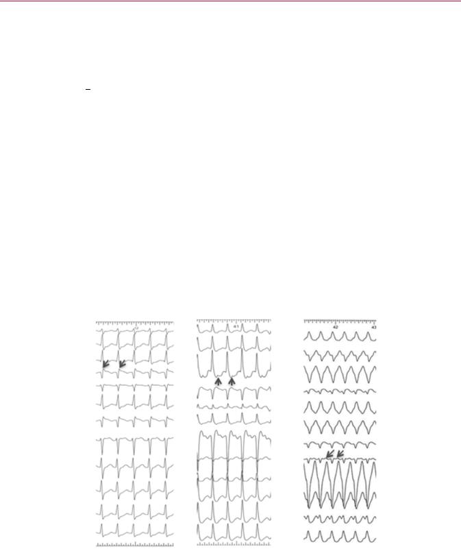
11.2.1.1.1 12 lead electrocardiogram during tachycardia. Typically, AVNRT is a narrow complex tachycardia, i.e. QRS duration <120 ms, unless there is aberrant conduction, which is usually of the RBBB type, or a previous conduction defect exists (Figure 13). AV dissociation is exceptionally uncommon, but it can occur as neither the atria nor the ventricles are necessary for the re-entry circuit. Thus, coexistence with AF or AV conduction block is possible but rare.66,322 ST-segment depression may be seen during or after the tachycardia.
In the typical form of AVNRT (also called slow fast AVNRT), retrograde P waves are constantly related to the QRS and, in the
A |
B |
C |
|
I |
|
|
|
II |
|
|
|
III |
|
|
|
aVR |
|
|
|
aVL |
|
|
|
aVF |
|
|
|
V1 |
|
|
|
V2 |
|
|
|
V3 |
|
|
|
V4 |
|
|
|
V5 |
|
|
|
V6 |
|
|
2019 |
|
|
|
|
|
25 mm/sec |
|
©ESC |
|
|
|
|
|
|
|
|
2019 September 13 on guest by 1093/eurheartj/ehz467/5556821.abstract/doi/10-article-com/eurheartj/advance.oup.https://academic from Downloaded
Figure 13 Atrioventricular nodal re-entrant tachycardia. (A) Typical atrioventricular nodal re-entrant tachycardia. (B) Atypical atrioventricular nodal reentrant tachycardia. (C) Atypical AVNRT with (unusual) left bundle branch block aberration. Retrograde P waves are indicated by arrows.

30 |
ESC Guidelines |
|
|
Table 12 Classification of atrioventricular nodal reentrant tachycardia types324
|
HA |
VA (His) |
AH/HA |
Typical AVNRT |
<70 ms |
<60 ms |
>1 |
Atypical AVNRT |
>70 ms |
>60 ms |
Variable |
|
|
|
|
Atypical atrioventricular nodal re-entrant tachycardia has been traditionally classified as fast slow (His atrial >70 ms, ventriculoatrial >60, atrial His/His atrial <1, and atrial His <200 ms) or slow slow (His atrial >70 ms, ventriculoatrial interval >60 ms, atrial His/His atrial >1, and atrial His >200 ms). Intermediate, unclassified types may also exist. AH = atrial His interval; AVNRT = atrioventricular nodal re-entrant tachycardia; HA = His atrial interval; VA = ventriculoatrial interval measured from the onset of ventricular activation on surface ECG to the earliest deflection of the atrial activation on the His bundle electrogram.
majority of cases, are indiscernible or very close to the QRS complex. Thus, P waves are either masked by the QRS complex or seen as a small terminal P’ wave that is not present during sinus rhythm.323
In the atypical form of AVNRT, P waves are clearly visible before
the QRS, i.e. RP>PR, denoting a long RP tachycardia, and are negative or shallow in leads II, III, aVF, and V6, but positive in V1.317
Tachycardia-related ST-segment depression, RR-interval variation, as well as QRS alternans may be seen. Specific, although modestly sensitive, ECG criteria for AVNRT, as opposed to AT and AVRT, are a pseudo R deflection in lead V1 and a pseudo S wave in the inferior leads, a notch in lead aVL, and a pseudo R in aVR.45 If the tachycardia is initiated by atrial ectopic beats, the initial (ectopic) P wave usually differs from the subsequent (retrograde) P waves.
11.2.1.1.2Electrophysiology study. Heterogeneity of both fast and slow conduction patterns has been well described, and all forms of
AVNRT may display anterior, posterior, and middle, or even LA retrograde, activation patterns.322 324 Thus, specific electrophysiologi-
cal manoeuvres may be required for differential diagnosis of typical
and, especially, atypical AVNRT from focal AT or AVRT due to a concealed septal pathway.45 The rare form of verapamil-sensitive AT is due to re-entry in the atrial tissue close to the AVN, but not the AV nodal conducting system.325
11.2.1.1.3Typical atrioventricular nodal re-entrant tachycardia. In the slow fast form of AVNRT, the onset of atrial activation appears before, at the onset of, or just after the QRS complex, thus maintaining an AH/His atrial (HA) ratio >1. The VA interval measured from the onset of ventricular activation on surface ECG to the earliest deflection of the atrial activation in the His bundle electrogram is <60 ms. Although, the earliest retrograde atrial activation is typically recorded at the His bundle electrogram, careful mapping studies
have demonstrated that posterior or even left septal fast pathways may occur in <7.6% in patients with typical AVNRT.326 328
11.2.1.1.4Atypical atrioventricular nodal re-entrant tachycardia.
Atypical AVNRT is seen in 6% of all AVNRT cases,317 and in some
.
.
.
.
.
.
.
.
.
.
.
.
.
.
.
.
.
.
.
.
.
.
.
.
.
.
.
.
.
.
.
.
.
.
.
.
.
.
.
.
.
.
.
.
.
.
.
.
.
.
.
.
.
.
.
.
.
.
.
.
.
.
.
.
.
.
.
.
.
.
.
.
.
.
.
.
.
.
.
.
.
.
.
.
.
.
.
.
.
.
.
.
patients may coexist with the typical form.329 A higher incidence of atypical AVNRT has been documented in athletes.330 In the socalled ‘fast slow form of AVNRT, retrograde atrial electrograms begin well after ventricular activation with an AH/HA ratio <1, indicating that retrograde conduction is slower than antegrade conduction. The AH interval is <185 200 ms. The VA interval measured from the onset of ventricular activation on surface ECG to the earliest deflection of the atrial activation in the His bundle electrogram is >60 ms. Earliest retrograde atrial activation has generally been reported at the base of the triangle of Koch, near the coronary sinus ostium, but it can be variable, with eccentric atrial
activation at the lower septum or even the distal coronary sinus.328,331,332 In the ‘slow slow’ form, the AH/HA ratio is >1 and the AH interval >200 ms, but the VA interval is >60 ms, suggesting that two slow pathways are utilized for both anterograde and retrograde activation. Earliest retrograde atrial activation is usually at the coronary sinus ostium, but variants of left-sided atrial retrograde activation have also been published.333,334 The distinction between ‘fast slow’ and ‘slow slow’ forms is of no practical significance, and certain cases of atypical AVNRT cannot be classified according to described criteria.324 There is also evidence that the ‘fast’ pathway during slow fast AVNRT is not identical to the ‘fast’ component of so-called fast slow AVNRT.329 Therefore, AVNRT can be classified as typical or atypical according to the HA interval, or—when a His bundle electrogram is not reliably recorded—according to the VA interval measured on the His bundle recording electrode.322 Table 12 presents a conventional classification system. Other approaches have also been published.335
11.2.1.2 Therapy
Recommendations for the management of atrioventricular nodal re-entrant tachycardia (AVNRT)
|
Recommendation |
Classa |
Levelb |
|
|
Acute therapy |
|
|
|
|
Haemodynamically unstable patients |
|
|
|
|
Synchronized DC cardioversion is recommended |
I |
B |
|
|
for haemodynamically unstable patients.86 88 |
|
||
|
|
|
|
|
|
Haemodynamically stable patients |
|
|
|
|
Vagal manoeuvres, preferably in the supine posi- |
I |
B |
|
|
tion with leg elevation, are recommended.41,89 91 |
|
||
|
|
|
|
|
|
Adenosine (6 18 mg i.v. bolus) is recommended |
I |
B |
|
|
if vagal manoeuvres fail.92 94 |
|
|
|
|
Verapamil or diltiazem i.v. should be considered if |
IIa |
B |
|
|
vagal manoeuvres and adenosine fail.92,94 98 |
|
||
|
|
|
|
|
|
Beta-blockers (i.v. esmolol or metoprolol) should |
|
|
|
|
be considered if vagal manoeuvres and adenosine |
IIa |
C |
|
|
fail.97,99,100 |
|
|
|
|
Synchronized DC cardioversion is recommended |
|
|
|
|
when drug therapy fails to convert or control the |
I |
B |
|
|
tachycardia.87,88 |
|
|
|
|
|
|
Continued |
|
2019 September 13 on guest by 1093/eurheartj/ehz467/5556821.abstract/doi/10-article-com/eurheartj/advance.oup.https://academic from Downloaded