Материал: Kaplan USMLE-1 (2013) - Anatomy
Section I • Histology and Cell Biology
myocardium and is composed mainly of cardiac muscle cells. The endocardium, which lines the chambers ofthe heart, is composed ofa simple squamous epithe lium, the endothelium, and a thin layer of connective tissue.
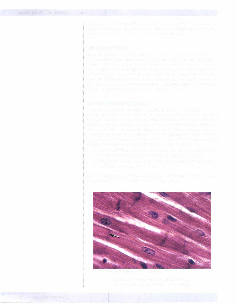
Intercalated Discs
Intercalated discs are special junctional complexes that join myocardial cells. The intercalated discs appear as dark, transverse lines in the light microscope. These disks contain gap junctions and adhering junctions. These junctions per mit the spread of electrical (gap) and mechanical (adhering) effects through the walls ofthe heart, synchronizing activity and for the pumping action ofthe heart chambers. While intercalated discs allow coordinated action of the myocardial cells, the squeezing and twisting movements of the heart chambers (particularly the left ventricle) during systole are due to the disposition of cardiac myocytes.
Cardiac Conduction System
The cardiac conduction system is a specialized group of myocardial cells that initiates the periodic contractions of the heart due to their ability to depolarize at a faster rate than other cardiac myocytes. Electrical activity spreads through the walls of the atria from the SA node and is quickly passed by way of internodal fibers to the atrioventricular node. From the atrioventricular node, activity pass es through the bundle of His and then down the right and left bundle branches in the interventricular septum. The bundle branches reach additional specialized cardiac muscle fibers known as Purkinje fibers in the ventricular walls.
•The Purkinje fibers run in several bundles along the endocardial surface and initiate ventricle activity starting at the apex of the ventricles.
•Purkinje fibers have a large cross section, a cytoplasm with few contrac tile fibrils and a large content of glycogen.
Cardiac muscle has a similar but somewhat less well developed T-tubule system compared to skeletal muscle that is located at the Z-line.
Copyright McGraw-Hill Companies. Used with permission.
Figure 1-4-6. Cardiac muscle cells with centrally placed nuclei and intercalated disks (arrow)
52 MEDICAL
Chapter If • Muscle Tissue
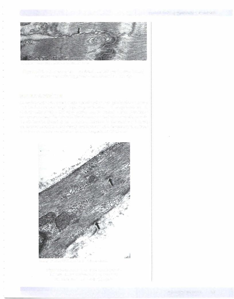
CopyrightMcGraw-Hill Companies. Used with permission.
Figure 1-4-7. EM of part of an intercalated disk with gap junction (arrow) adjacent to an adhering junction with intracellular density.
SMOOTH MUSCLE
Smooth-muscle cells have a single central nucleus, are spindle-shaped, have a variable diameter and length depending on location, and are generally smaller than the cells of either skeletal or cardiac muscle. Smooth muscle often forms one or more layers ofhollow tube-like structures in the body. Generally, smooth muscle bundles (fascicles} run in various directions in the bladder and ureter, are circumferential in blood vessels, and form at least 2 discrete layers, an inner circular and an outer longitudinal layer, in the gastrointestinal tract.
Copyright McGraw-Hill Companies. Used with permission.
Figure 1-4-8. EM of part of a smooth-muscle cell with dense bodies (arrows) that form attachment sites foractin filaments
MEDICAL 53
Section I • Histology and Cell Biology
Smooth muscle is not striated because it lacks the sarcomeres and Z lines seen in striated muscle. Instead, the actin filaments are oriented in multiple oblique, roughly longitudinal, directions and are anchored to the inner cell membrane at multiple sites, not just at the ends of the cells (the arrangement in striated muscle). This allows more shortening of smooth-muscle cells compared to stri ated muscle. Instead ofaligned Z bands, the actin filaments are anchored in dense bodies scattered within the cell cytoplasm. The dense bodies also anchor the con tractile filaments to the cell surface membrane. In vascular smooth muscle, the dense bodies contain the intermediate filaments desmin and vimentin. In smooth muscle of the gastrointestinal tract, the dense bodies contain desmin but not vi mentin. Smooth muscle lacks a T-tubular system, and the SR has a less ordered relationship to contractile fibers.
ChapterSummary
• Muscles are classified as skeletal, cardiac, or smooth.
Skeletal Muscle
•Skeletal muscle has 3 levels of connective tissue: endomysium, perimysium,
and epimysium. Skeletal muscle is composed of long cylindrical fibers that have dark (A) bands and light (I) bands. A dark transverse line, the Z line, bisects each I band. Skeletal muscle fibers contain myofibrils, which in turn are composed of sarcomeres.
•Sarcomeres have thick and thin filaments. Thick filaments are centrally located in sarcomeres, where they interdigitate with thin filaments. The I band contains thin filaments only, the H band contains thick filaments only, and the A bands contain both thick and thin filaments.
•Thin filaments contain 3 proteins: actin, tropomyosin, and troponin.
•Actin forms a double helix, whereas tropomyosin forms an a-helix. Troponin includes 3 polypeptides: TnT, which binds to tropomyosin; TnC, which binds to calcium ions; and Tnl, which inhibits actin-myosin interaction. Thick filaments are composed of myosin. Myosin has 2 heavy chains with globular head regions. The heads contain actin-binding sites and have ATPase activity. The transverse tubular system (T-tubule system) surrounds each myofibril and facilitates excitation-contraction coupling.
Cardiac Muscle
•Cardiac muscle has an arrangement of sarcomeres similar to that in skeletal muscles, but the fibers are coupled through gap junctions.
•Smooth muscle is found in the walls of blood vessels and hollow viscera.
Gap junctions couple them electrically. Myofilaments of smooth muscles are not arrayed like in skeletal muscles; they are obliquely placed in order to form a latticework. Electrical or chemical signaling via hormones can trigger smooth muscles.
54 M EDICAL

Nervous Tissue |
5 |
NEURONS
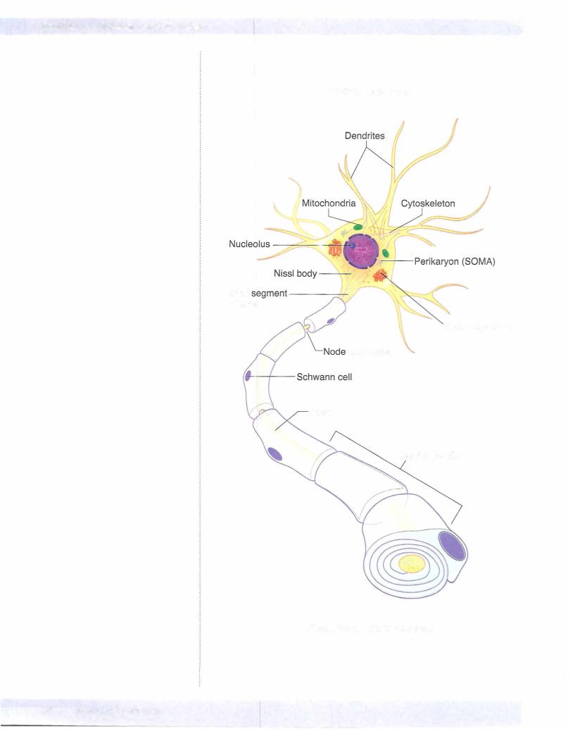
Neurons are cells that are morphologically and functionally polarized so that information may pass from one end ofthe cell to the other.
Neurons may be classified by the form and number oftheir processes as bipolar, unipolar, or multipolar. The cell body of the neuron contains the nucleus and membrane-bound cytoplasmic organelles typical of a eukaryotic cell, including endoplasmic reticulum (ER), Golgi apparatus, mitochondria, and lysosomes. The nucleus and nucleolus are prominent in neurons.
The cytoplasm contains Nissl substance, clumps ofrough ER with bound poly somes. The cytoplasm also contains free polysomes; free and bound polysomes in the Nissl substance are sites ofprotein synthesis.
MEDICAL 55