Материал: Kaplan USMLE-1 (2013) - Anatomy
Chapter 5 • Nervous Tissue
CopyrightMcGraw-Hill Companies. Used withpermission.
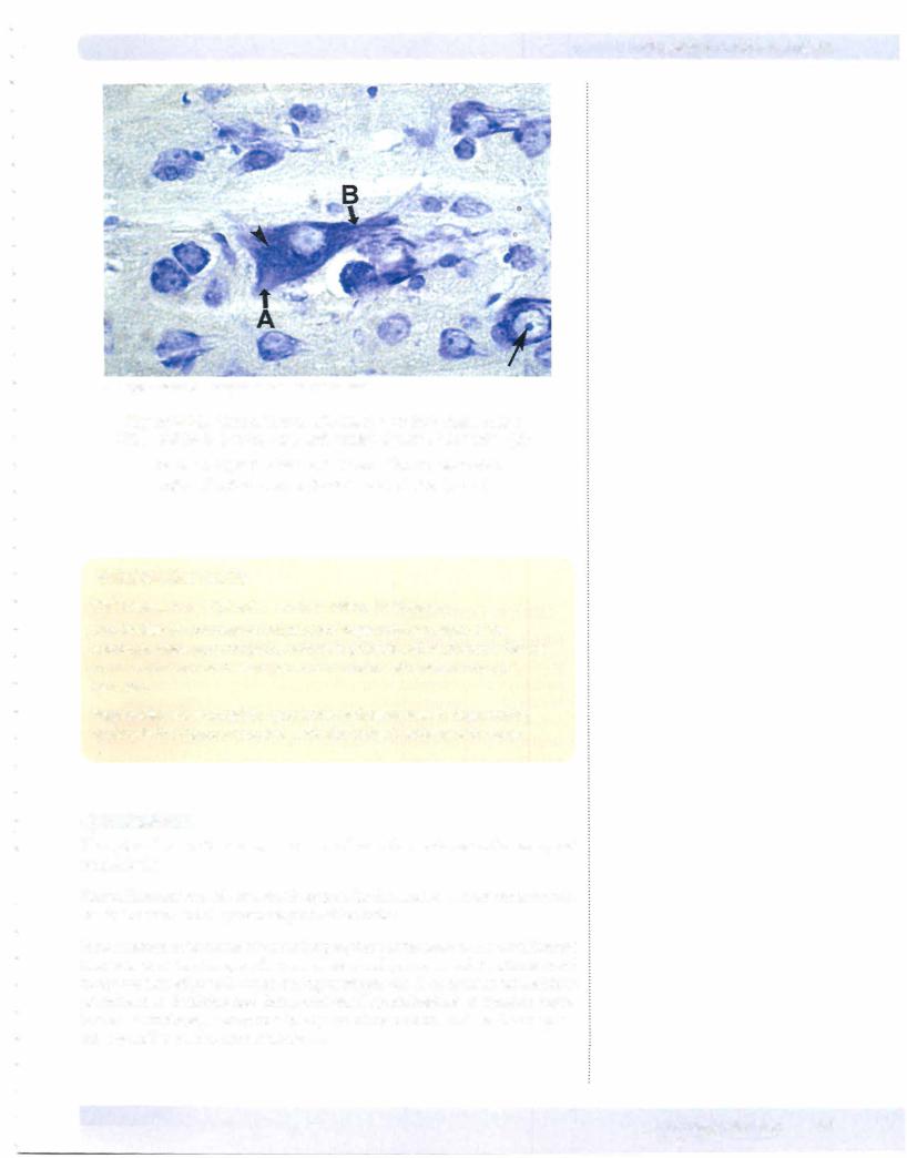
Figure 1-5-2. Neural Tissue with Nissl stain that stains rough
ER in cell body (arrowhead) and proximal parts of dendrites (B)
The axon (A) lacks Nissl substance. The nucleus of an adjacent neuron has a prominent nucleolus (arrow).
Clinical Correlate
CNS Disease and Cytoplasmic Inclusions in Neurons
Lewy bodies are cytoplasmic inclusions of degenerating neurons ofthe substantia nigra, pars compacta, evident in patients with Parkinson's disease and in cortical and brain-stem neurons in patients with certain forms of dementia.
Negri bodies are eosinophilic cytoplasmic inclusions seen in degenerating neurons in the hippocampus and cerebellar cortex in patients with rabies.
Cytoskeleton
The cytoskeleton ofthe neuron consists ofmicrofilaments, neurofilaments, and microtubules.
Neurofilaments provide structural support for the neuron and are most numer ous in the axon and the proximal parts ofdendrites.
Microfilamentsform a matrixnear the peripheryofthe neuron. A microfilament matrix is prominent in growth cones ofneuronal processes and functions to aid in the motility ofgrowth cones during development. A microfilament matrix is prominent in dendrites and forms structural specializations at synaptic mem branes. Microtubules are found in allparts ofthe neuron, and are the cytoplas mic organelles used in axonal transport.
MEDICAL 57
Section I • Hi tology and Cell Biology
Copyright McGraw-Hill Companies. Used with permission.
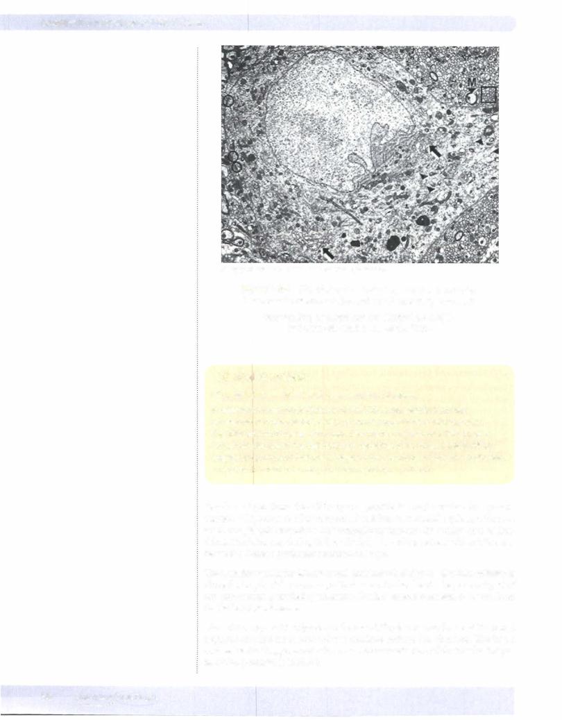
Figure 1-5-3. EM of Neuropil including the Cell Body of a
Neuron with Rough ER (arrowheads) and Golgi (arrows)
Surrounding neuropil has myelinated axons(M) and unmyelinated bare axons (box).
Clinical Correlate
Microtubules and Neuronal Degenerative Diseases
In degenerative neuronal diseases of the CNS, a tau protein becomes excessively phosphorylated, which prevents crosslinking of microtubules. The affected microtubules form helical filaments and neurofibrillary tangles and senile plaques in the cell body and dendrites of neurons. Neurofibrillary
tangles are prominent features of degenerating neurons in Alzheimer's disease, amyotrophic lateral sclerosis, and Down syndrome patients.
Dendrites taper from the cell body and provide the major surface for synaptic contacts with axons of other neurons. which are small cytoplasmic extensions that dramatically increase the surface area of den drites. Dendrites maybe highlybranched; thebranchingpattern ofdendrites may be used to define a particular neuronal cell type.
Theaxonhas auniformdiameterandmaybranchatrightanglesinto collaterals along the length ofthe axon, in particular near its distal end. The proximal part of the axon is usually marked by an a tapered extension ofthe cellbody that lacks Nissl substance.
The initial segment is adjacent to the axon hillock. The membrane ofthe initial segment contains numerous voltage-sensitive sodium ion channels. The initial segment is the "trigger zone" ofan axon where conduction ofelectrical activity as an action potential is initiated.
58 MEDICAL


Section I • Histology and Cell Biology
remove the neurotransmitter glutamate and K+ ions from the extracellular space. Astrocytes have foot processes that contribute to the blood-brain bar rier by forming a glial-limiting membrane. Astrocytes hypertrophy and pro liferate after an injury to the CNS; they fillup the extracellular space left by degenerating neurons by forming an astroglial scar. Radial glia are precursors of astrocytes that guide neuroblast migration during CNS development.
Microglia cells are the smallest glial cells in the CNS. Unlike the rest of the CNS neurons and glia, which are derived from neuroectoderm, microglia are derived from bone marrow monocytes and enter the CNS after birth. Microglia provide a link between cells of the CNS and the immune system. Microglia proliferate and migrate to the site of a CNS injury and phagocytose neuronal debris after injury. Pericytes are microglia that contribute to the blood-brain barrier.
GLIAL CELLS
Microglia and CNS Disorders
Microglia determine the chances of survival of a C S tissue graft and are the cells in the CNS that are targeted by the HIV- 1 virus in patients with AIDS. The affected microglia may produce cytokines that are toxic to neurons.
CNS microglia that become phagocytic in response to neuronal tissue damage may secrete toxic free radicals. Accumulation offree radicals, such as superoxide, may lead to disruption of the calcium homeostasis of neurons.
Oligodendrocytes form myelin for axons in the CNS. Each of the processes of the oligodendrocyte can myelinate individual segments of many axons. Unmy elinated axons in the CNS are not ensheathed by oligodendrocyte cytoplasm.
Schwann cells are the supporting cells of the peripheral nervous system (PNS), and are derived from neural crest cells. Schwann cells form the myelin for axons and processes in the PNS. Each Schwann cell forms myelin for only a single inter nodal segment of a single axon. Unmyelinated axons in the PNS are enveloped by the cytoplasmic processes of a Schwann cell. Schwann cells act as phagocytes and remove neuronal debris in the PNS after injury. A node ofRanvier is the region between adjacent myelinated segments of axons in the CNS and the PNS.
In all myelinated axons, nodes ofRanvier are sites that permit action potentials to jump from node to node (saltatory conduction). Saltatory conduction dramati cally increases the conduction velocity of impulses in myelinated axons.
60 MEDICAL
Chapter 5 • NervousTissue
CopyrightMcGraw-Hill Companies. Used with permission.
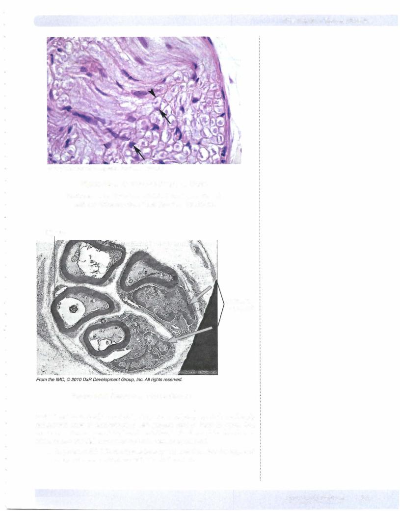
Figure 1-5-4. Section of a Peripheral Nerve
Endoneurial border of an individual axon (arrowhead) and two Schwann cell nuclei (arrows) are shown.
Myelin
Schwann cell nuclei
Figure 1-5-5. Axons Cut in Cross-Section
Multiple sclerosis (MS) is marked by the presence of plaques, which are sharply demarcated areas of demyelination. MS plaques tend to form in axons that course near the surfaces of the lateral ventricles, in the floor of the fourth ven tricle, or near the pial surfaces of the brain stem or spinal cord.
•In patients with MS, multiple lesions appear over time, but the signs and symptoms may undergo exacerbation and remission
MEDICAL 61