Материал: Kaplan USMLE-1 (2013) - Anatomy
Section I • Histology and Cell Biology
Both thetrabeculae ofnewlyformed woven bone and the newlyformed densebone oftheoutercortexgth undergoremodeling, the end result ofwhichis to produce high ly ordered larnellae ofcollagen in the matrix, which gives the mineralized osteoid greater stren and resistance to breaking. This forms lamellar or cortical bone.
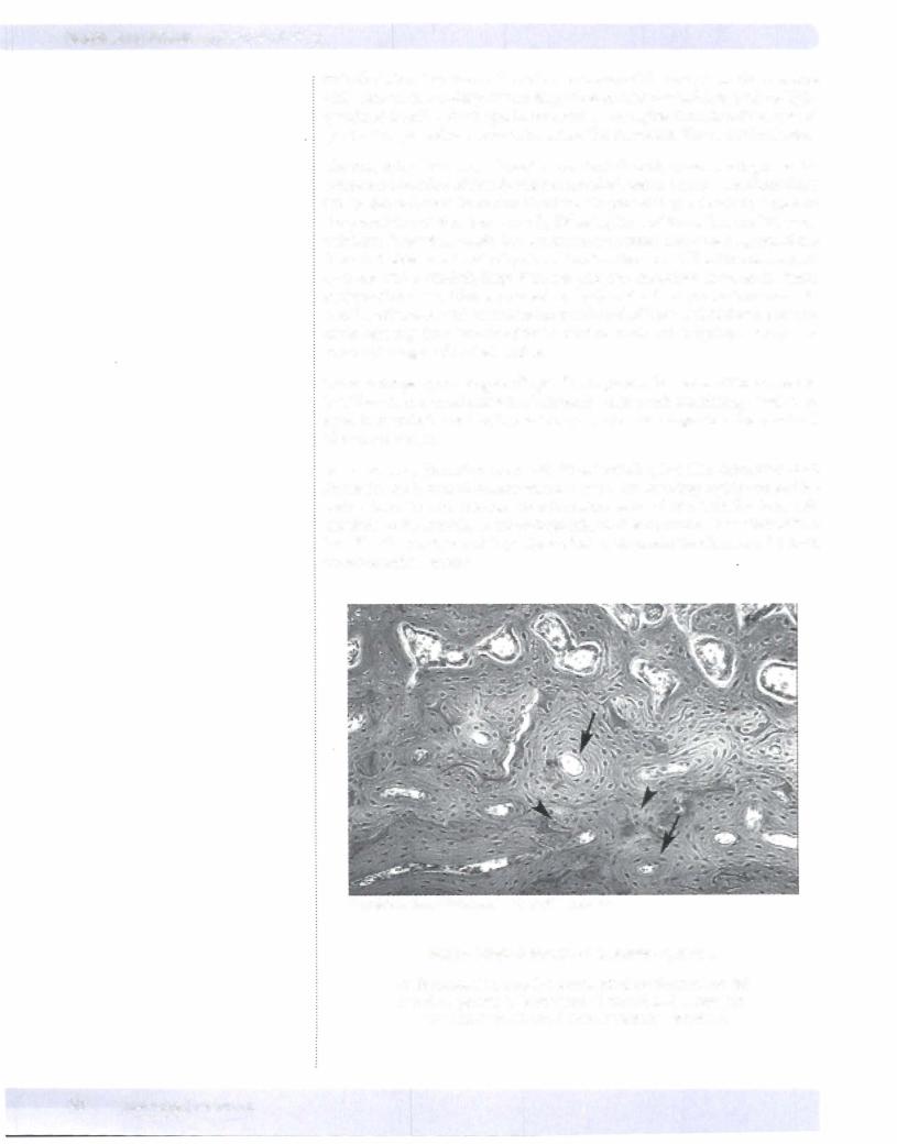
The processbywhich wovenbone isconvertedtolamellarbone is analogous to the constant remodeling ofalready existing lamellar bone that takes place throughout life. In dense cortical bone new blood vessels, preceded by osteoclasts, bore into the preexisting cortical bone, usually following lines of stress. The resulting tun nels form Haversian canals that are generally oriented along the long axis ofthe bone, but otherwise directed by stress lines in other bones. The Haversian canals start out with a relatively large diameter, and new osteoblasts form on the inner surface ofthe osteoid thatwascarved outbythe osteoclasts. Successive concentric lamellae ofosteoidwith alternating orientation ofcollagen are laid down, progres sively reducing the diameter of the Haversian canals and trapping osteocytes in concentric rings within the lamellae.
There is always a persisting small cylindrical space in the center ofthe concentric lamellae where a small amount ofconnective tissue with nourishing vessels per sists. Even at their smallest, Haversian canals are muchlarger than the canaliculi that connect them.
An osteon is a Haversian canal with its surrounding lamellae constituents and forms the basic unit of mature lamellar bone. The resulting highly ordered la mellar bone is much stronger than immature woven bone. Lamellar bone will continue to be remodeled throughout life, so it is common to see interstitial lamellae that are incomplete portions ofolder osteons located between the more recent complete osteons.
Copyright McGraw-Hill Companies. Used with permission.
Figure 1-3-8. Osteons or Haversian systems
Each Haversian canal is surrounded by circumferential lamellae (arrows). Remnants of remodeled Haversian systems (arrowheads) form interstitial lamellae.
42 MEDICAL
Chapter 3 • cartilage and Bone
Trabeculae of cancellous bone are similarly remodeled. The process takes place mostly at the surface of the trabeculae, carried out by osteoclasts and osteoblasts from the adjacent marrow.
Mature long bones are covered on all surfaces by dense cortical bone with an outer periosteum, with the exception of articular surfaces, which are covered by hyaline articular cartilage. The inner bone at the epiphyses and the nearby end portions of the diaphyses (the flared metaphyses) are filled with cancellous bone and bone marrow. The central most portion ofthe diaphysis oflarger long bones is filled with bone marrow, either hematopoietically active or fatty, without inter vening bony trabeculae.
The main nutrient arteries ofa long bone remain as relatively large arteries enter ing the diaphysis and the epiphyses, Once in the medullary cavity, they give rise to many small branches, some of which can reenter the cortex from the inner surface through Volkmann's canals. In addition there are many small branches along the periosteum, which in turn may perforate into the cortical bone from the outside, also giving rise to additional Volkmann's canals, all ofwhich are per pendicular to Haversian canals, Volkmann's canals are perpendicular to Haver sian canals, and are distinguished by the lack of a surrounding osteon.
MEDICAL 43
Section I • Histology and Cell Biology
ChapterSummary
•Cartilage like all connective tissue, consists of cells and extracellular matrix (ECM).
•Cartilage formation takes place by differentiation of multipotential mesenchymal cells into chondroid precursor cells, which give rise to chondroblasts, which give rise to chondrocytes. Cartilage can enlarge in 2 ways. In appositional growth, new cells can be added from the outer
perichondrium. In internal or interstitial growth, the chondrocytes embedded deep within the cartilage can continue to produce additional ECM. There are 3 types of cartilage, all containing type II collagen and glycosaminoglycans (GAGs), but sometimes with additional extracellular components, which were produced by the chondrocytes.
•Hyaline cartilage is the type of cartilage that forms a template for bone formation during embryogenesis, as well as comprising the cartilage on the surface of bones at synovial joints and the cartilage of the nose, portions of the larynx, and the cartilage ofthe trachea and bronchi. Elastic cartilage is found in the external ear, the auditory canal, and the epiglottis ofthe larynx. Fibrocartilage is the type of cartilage found in intervertebral disks ofthe vertebral column and the menisci ofthe knee, and may form the attachment of ligaments and tendons to bone.
•
•
Bone is a unique connective tissue in that it not only has cells and ECM called osteoid, including type I collagen and GAGs, but also the matrix is calcified and rigid.
Osteoblasts secrete bone extracellular matrix (osteoid). Osteoblasts are specialized to synthesize and secrete the components of osteoid, type I collagen, and GAGs.
•Osteoclasts are responsible for the breakdown of bone matrix, with release of calcium.
•All bone, regardless offormation, undergoes remodeling throughout life. All 3 processes-bone formation, bone growth, and bone remodeling-have similarities.
•In intramembranous bone formation, primitive mesenchyme can give rise directly to bone. In endochondral bone formation, bone is formed on the template of preexisting hyaline cartilage. Endochondral bone formation occurs in long bones ofthe extremities and vertebrae and bones ofthe pelvis, and starts as a hyaline cartilaginous template.
•An osteon is a Haversian canal with its surrounding lamellae constituents and forms the basic unit of mature lamellar bone. Trabeculae of cancellous bone are similarly remodeled. The process takes place mostly at the surface of the trabeculae, carried out by osteoclasts and osteoblasts from the adjacent marrow.
44 MEDICAL