Материал: Kaplan USMLE-1 (2013) - Anatomy
Section I • Histology and Cell Biology
Table 1-4-1. Red versus White Skeletal Muscle Fibers
Red Fibers (Type I)
Slow contraction
.J, ATPase activity
i Capacity for aerobic metabolism
iMitochondrial content
iMyoglobin (imparts red color)
Best for slow, posture-maintaining muscles, e.g., back (think chicken drumstick/thigh)
White Fibers (Type II)
Fast contraction i ATPase activity
i Capacity for anaerobic glycolysis
.J, Mitochondrial content
.J, Myoglobin
Best for fast, short-termed, skilled motions, e.g., extraocu lar muscles of eye, sprinter's legs, hands (think chicken breast meat and wings)
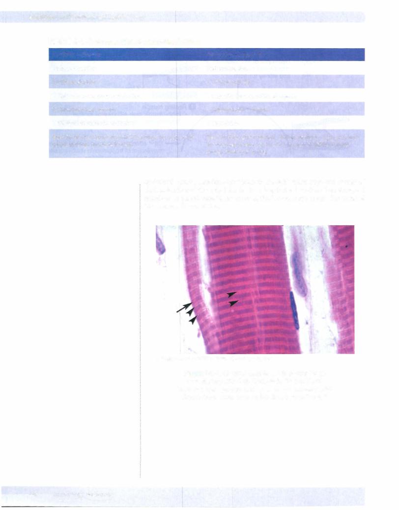
In skeletal muscle, striations are visible in the light microscope and consist of dark A bands and the light I bands. In a longitudinal section of an elongated muscle cell a pale H band in the center of the A band may be seen. 'Ihe Z and M lines are usually not visible.
Copyright McGraw-Hill Companies. Used with permission.
Figure 1-4-4. Skeletal muscle cell that consists of sarcomeres with dark A bands in the middle of
the sarcomere (arrow) and light l bands (arrowheads) A peripheral nucleus is on the right side of the cell.
48 MEDICAL
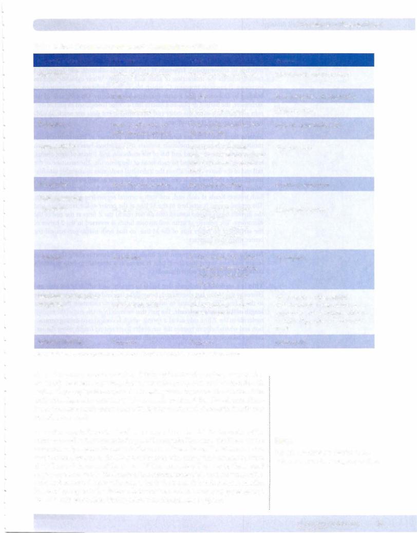
Table 1-4-2. Structure, Function, and Pharmacology of Muscle
Characteristics
Appearance
Ttubules
Cell junctions
Innervation
Action potential
Upstroke
Plateau
Excitation-contraction coupling
Calcium binding
Skeletal
Striated, unbranched fibers
Z lines
Multinucleated
Form triadic contacts with SR at A-I junction
Absent
Each fiber innervated
Inward Na+ current
No plateau
AP T tubules Ca2+ released from SR
Troponin
Cardiac
Striated, branched fibers
Z lines
Single central nucleus
Form dyadic contacts with SR near Z line
Junctional complexes between fibers (intercalated discs), including gap junc- tions
Electrical syncytium
•Inward Ca2+ current (SA node)
•Inward Na+ current (atria, ventricles, Purkinje fibers)
•No plateau (SA node)
•Plateau present (atria, ventricles, Purkinje fibers)
Inward Ca2+ current during plateau Ca2+ release from SR
Troponin
Abbreviations: AP, action potential; IP3, inositol triphosphate; S R, sarcoplasmic reticulum
Chapter 4 • Muscle Tissue
Smooth
Nonstriated, fusiform fibers
No Z lines; have dense bodies
Single nucleus
Absent; have limited SR
Gap junctions
Electrical syncytium
Inward Na+ current
No plateau
AP opens voltage-gated Ca2+ channels in sarcolemma;
hormones and neurotransmittersopen IP3-gated Ca2+ channels in SR
Calmodulin
Anendomysium surrounds each skeletal muscle cell and provides insulation be tween adjacent cells. A perimysium surrounds a group or fascicle ofmuscle cells and is where capillaries are found. The epimysium surrounds the outside ofthe entire muscle, and is where larger bloodvessels are found. Satellite cells and fibro blasts that have small dark nuclei with tightly condensed chromatin lie adjacent to each muscle cell.
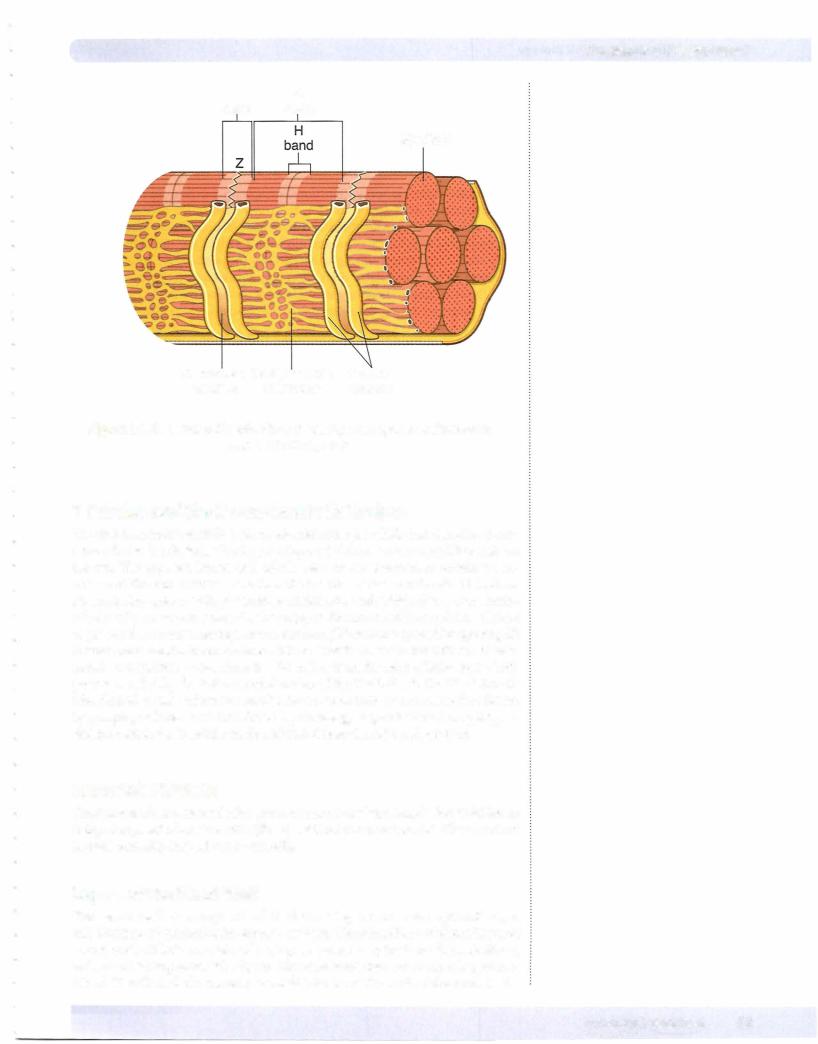
The Z lines mark the ends ofeach sarcomere. The A band is in the center ofthe sarcomere and is the zone including all ofthe myosin filaments. The H zone is the zone with only myosin filaments in the center ofthe A band. The M line is in the center ofthe sarcomere. The I band is the zonewith only actin filaments between the Z line and the start ofthe A zone. When a muscle cell contracts, the A band stays constant in width (the length ofthe myosin molecule). As the overlap ofthe actin and myosin filaments increases, both the I and H bands become smaller. The overlap region is the distance between the I and H bands, and grows as the I and H bands get smaller. During relaxation, the opposite happens.
Note
Desmin intermediate filaments link adjacent myofibrils at adjacent Z lines.
MEDICAL 49
Section I • Histology and Cell Biology
Clinical Correlate
Skeletal muscle has a limited ability to regenerate. Satellite cells are stem cells situated outside the plasma membrane that form new myoblasts.
Muscular dystrophies deplete the pool of satellite cells.
Actin thin filaments in muscle consist of 2 strands of F-actin forming a helix, around which tropomyosin filaments are wrapped. Tropomyosin serves as a site of attachment for troponin, which has 3 component parts: (which attaches to tropomyosin), troponin C (which binds calcium), and troponin I (which inhibits the interaction of actin and myosin). When cytosolic calcium increases, it binds to troponin C, which blocks the inhibition of actin/myosin binding by troponin I. The F-actin filaments have polarity. On each side of the sarcomere, the barbed or plus end inserts into a Z line (composed in part ofdes min and actinin), while the other end faces toward, but does not reach, the M line in the center ofthe sarcomere.
Myosin thick filaments are formed of a bipolar polymer of individual myosin molecules. Each myosin molecule consists of 2 identical heavy chains, which each contribute to the head and tail ofthe molecule, and 2 pairs oflight chains, with one ofeach type bound to each head ofthe molecule. The structure ofthe tail end ofthe heavy chains allows the individual molecules to assemble into fila ments, while the head ends can interact with myosin as well as hydrolyze ATP. When the myosin molecules self-assemble, they form a bipolar thick filament, with myosin heads at each end, and with a central region free ofmyosin heads. The central region is attached to the M line at the center ofthe sarcomere, and the myosin heads extend toward (but do not reach) the Z lines at the end ofthe sarcomere. The polarity ofactin and myosin heads is reversed in the 2 halves of the sarcomere on either side of the M line, so that both halves pull toward the centerwhen they contract.
Nebulin is a scaffolding protein that binds to actin filaments along their entire length and also insert into the Z line. It serves as a template to help maintain constant length ofthe actin filaments.
Titin is a scaffolding molecule that binds to myosin, and which is not only anchored in the M line but also extends beyond the free end ofthe myosin molecule to the Z line. The part bound to myosin again helps to maintain the constant lengthofthe myosin molecule. Thepartthat extends from the end ofthe myosin molecule to the Z line may act as a spring, which is compressed during contrac tion and which helps to restore the sarcomere to its resting length when the sar comere relaxes, at the end ofactin/myosin interaction.
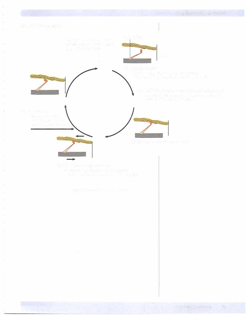
Contractions ofskeletal muscle produce force and movement by the interaction of thin actin (F-actin) and thick myosin (myosin II) filaments. The interaction uses energyderived fromATP and involves repetitive binding, sliding, release and reattachment ofthe head end of the myosin molecules to the adjacent actin fila ments. The contraction is ultimately startedby releasing calcium, which promotes binding and cocking ofthe myosin head, hydrolysis ofATP and movement ofthe myosin head back to its starting position. Relaxation ofskeletal muscle involves a reduction ofcytosolic calcium to halt the interaction ofmyosin and actin.
50 MEDICAL
Chapter 4 • Muscle Tissue
I A band band
Myofibril
Transverse Sarcoplasmic Terminal
tubules reticulum cisterna
Figure 1-4-5. Striated Muscle Fiber Showing Sarcoplasmic Reticulum
and T-tubule System
T Tubules and the Sarcoplasmic Reticulum
The plasma membrane ofskeletalmuscle cells extendsinfolds into the cell as trans verse tubules (T tubules), allowing rapid spread ofthe action potential throughout the cell. The apparent lumen of T tubules seen in cross section is actually an ex tension of the extracellular space into the interior of the muscle cell. The action potential also opens a voltage-sensitive calcium channel, which allows some extra cellular calcium to enter the cell. This triggers the release of intracellular calcium sequesteredin adjacentsarcoplasmicreticulum (SR) intothe cytosolbyopeningSR membrane-located calcium channels. The bulk ofthe calcium that initiates skeletal muscle contraction comes from the SR rather than the extracellular space. This process is aided by the intimate relationship ofthe T tubules to the SR at the tri ads. Skeletal muscle relaxation results from a reduction of free cytosolic calcium by pumping calcium back into the SR by an energy-dependent calcium pump. In skeletal muscle the T-tubule triads are located near the A-I band junction.
CARDIAC MUSCLE
Cardiac muscle is striated in the same manner as skeletal muscle, but it differs in being composed ofsmaller cells (fibers) with only one or 2 nuclei. The nuclei are located centrally, instead ofperipherally.
Layers ofthe HeartWall
The heart wall is composed of 3 distinct layers: an outer epicardium, a middlemyocardiumandaninnerendocardium.Theepicardium,orviscerallayerof serous pericardium, consists of a simple squamous epithelium (mesothelium) and its underlyingconnective tissue. The connective tissue contains a large num ber of fat cells and the coronary vessels. The muscular wall of the heart is the
MEDICAL 51