Материал: Kaplan USMLE-1 (2013) - Anatomy

Chapter 6 • Immune Tissues
Lymph Circulation
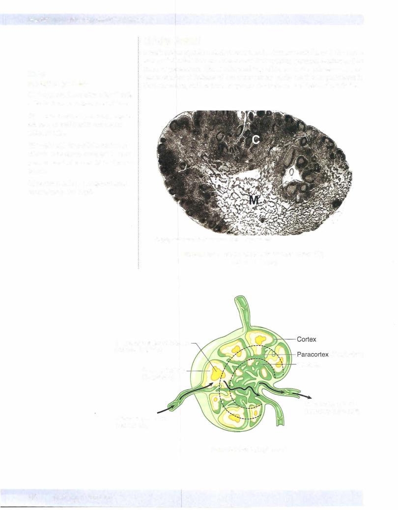
In lymph nodes, the B and T cells have exited the blood circulation, having come initially from bone marrow or thymus, respectively, but continue to circulate from one site to another for long periods, up to years. The lymph nodes also contain antigen-presenting cells and provide opportunities for lymphocytes to encounter antigens that they have been programmed to recognize, either for the first time or after prior encounter with resulting proliferation, maturation, and migration from another part ofthe immune system.
Afferent lymphatics carry lymph from peripheral tissues or other lymph nodes. Afferent lymphatics enter lymph nodes bypiercing the capsule ofthe node around its periphery. The lymph drains into a peripheral which empties into smaller paratrabecular sinuses that pass through the cortex ofthe node. In the cortex are resident T and B lymphocytes, macrophages and antigen presenting cells, and stromalreticulin cells, as well as smallblood vessels, includingpost-cap illaryvenules withhigh (cuboidalrather thansquamous) endothelialcells
HEVs are specialized to allow lymphocytes to exit the bloodstream in nodes or other extranodal lymphoid sites.
Efferent lymphatics convey T-cell progeny and B memory cells from the node at the medulla at the hilum ofthe node and travel via lymphatics to other lymph
nodes or the bloodstream. Some B cells give rise to plasma cells that populate the medullary cords deep to the cortex and secrete antibody into blood and lymph, whichis then carried to the rest ofthebody. Normallyfewifanyplasmacells circu late in the peripheral blood, in contrast to other lymphocyte cell types.
MUCOSA-ASSOCIATED LYMPHOID TISSUE OF THE GUT (GALT)
Inmucosa-associatedlymphoid tissue ofthe gut (GALT) the overlying epithe lium can be specialized to transport antigens through the epithelial layer to the underlying lymphoid cells. The ileum has a high concentration of GALT orga nized into Peyer's patches, which are partially covered on their luminal surface by special microfold or M cells. M cells are epithelial cells that can pick up anti gens from the ileal lumen and present them to B cells, which have surface immu noglobulin that recognizes the antigens. The B cells that recognize antigen from the M cells then present the antigen to T-helper cells via follicular dendritic cells.
MEDICAL 69
Section I • Histology and Cell Biology
Copyright McGraw-Hi/I Companies. Used with permission.
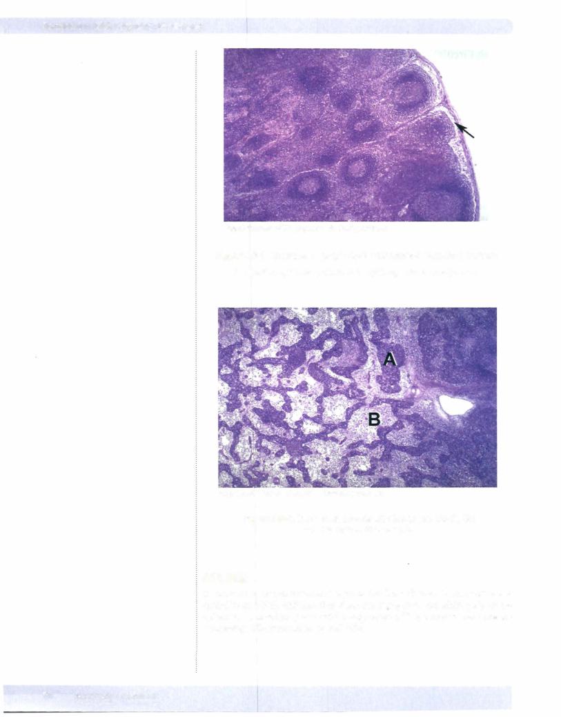
Figure 1-6-3. Cortex of a Lymph Node with Several Germinal Centers
Afferent lymphatics drain into the subcapsular sinus (arrow).
Copyright McGraw-HillCompanies. Used with permission.
Figure 1-6-4. Medulla of a Node with Medullary Cords (A) and Medullary Sinuses (8)
SPLEEN
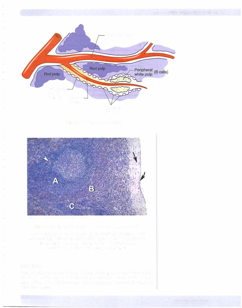
In contrast to lymph nodes and thymus that have distinct layers (cortex and medulla) in which different functions are segregated, the white pulp of the spleen is arranged in periarteriolar aggregates of lymphocytes and antigen processing cells surrounded by red pulp.
70 MEDICAL
Chapter 6 • Immune Tissues
Trabecular artery
Periarterial sheath
(T cells) Central artery
Marginal
Sinuses
Figure 1-6-5. Spleen Schematic
CopyrightMcGraw-Hill Companies. Used with permission.
Figure 1-6-6. Spleen that lacks a distinct cortex and medulla.
Areas of white pulp (A) are randomly interspersed with red pulp (B) and connective tissue trabeculae (C). There is a dense connective tissue capsule (arrow), a covering layer of mesothelium
(curved arrow) and central artery (arrowhead).
Red Pulp
Red pulp filters and clears the bloodstream ofdamaged or aging red blood cells, particulate matter, large antigens including bacteria and unwanted cells. It carries this function out in a distinct stroma with macrophages. The red pulp consists of cords and sinuses.
MEDICAL 71