Материал: Kaplan USMLE-1 (2013) - Anatomy

Respiratory System |
7 |
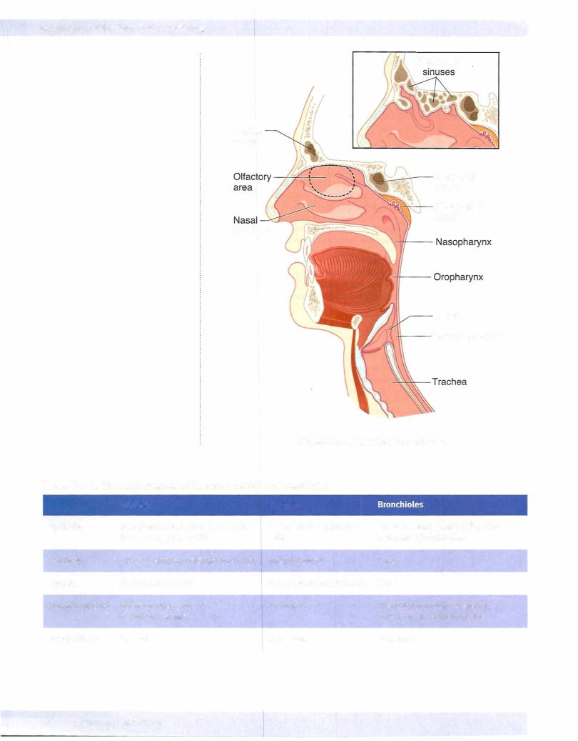
The lung is an organ that functions in the intake ofoxygen and exhaling of C02. Approximately 14 times each minute, we take in about 500 ml ofair per breath. Inspired air willbe spread over 120 square meters ofthe surface area ofthe lungs. The air-blood barrier has to be thin enough for air to pass across but tough enough to keep the blood cells inside their capillaries.
Lungs are opened to the outside world so that they are susceptible to environ mental insults in the form ofpollution and infectious bacteria.
The lungs receive the entire cardiac output and are positioned to modify various blood components. The pulmonary endothelium plays an active role in the meta bolic transformation of lipoproteins and prostaglandins. The enzyme that con verts angiotensin I to angiotensin II is produced bythe lung endothelial cells.
Clinical Correlate
•Any disease that affects capillaries also affects the extensive capillary bed ofthe lungs. Bacteria which colonize the lungs may damage the barriers between the alveoli and the capillaries, gaining access to the bloodstream (a common complication of bacterial pneumonia).
•In individuals with allergies, smooth-muscle constriction reduces the diameter of air tubes and results in reduced air intake.
•Lung cancers commonly develop from bronchi (smoking, asbestos, and excessive radiation are the main causes).
•Mesothelioma is a malignant tumor of the pleura (causative agent: asbestos dust).
MEDICAL 77
Chapter 7 • Respiratory System
PULMONARY DEFENSE MECHANISMS
Inhaled air contains hazardous dusts, chemicals, and microorganisms, yet lung tissue is usually sterile. Inhaled particles are deposited on the walls of airways according to size.
•Particles 10 microns in diameter or larger are trapped in the nose and are removed by sneezing, blowing, or swallowing.
•Particles 3-10 microns in diameter are trapped and removed by the tracheobronchial mucociliary escalator.
•Particles 0.5-3 microns in diameter (bacterial size) may reach the alveolar tissue and are removed by alveolar clearance.
•Macrophages that reside in the alveolar spaces phagocytose the small particles and transport them by amoeboid movement into bronchioles, lymphatics, and capillaries. Sometimes they remain trapped in connec tive tissue septa.
TRACHEA
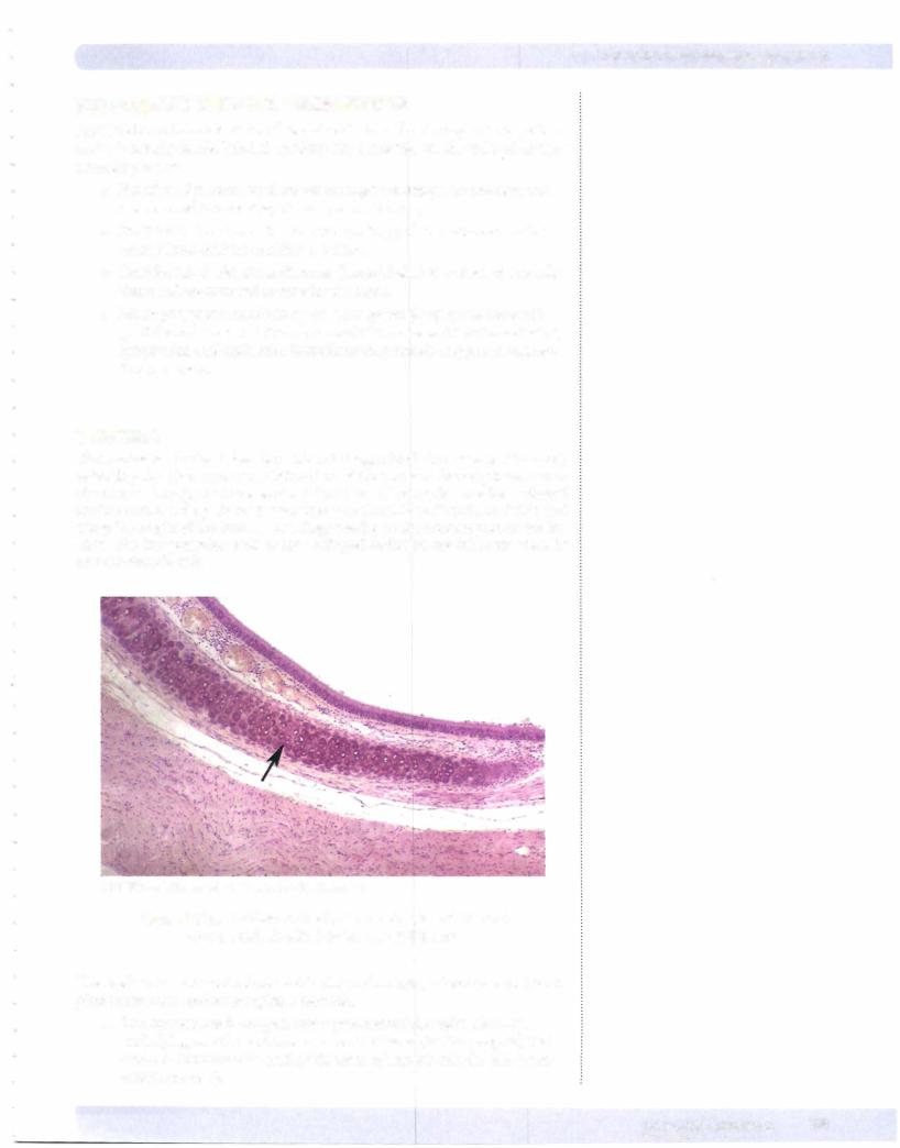
The trachea is a hollow tube, about 10 cm in length (and about 2 cm in diameter), extending from the larynx to its bifurcation at the carina to form a primary bron chus for each lung. The most striking structures of the trachea are the C-shaped hyaline cartilage rings. In the human there are about 16 to 20 ofthem distributed along the length of the trachea. The rings overlap in the anterior part of the tra chea. The free posterior ends of the C-shaped cartilages are interconnected by smooth-muscle cells.
Copyright McGraw-Hill Companies. Used with permission.
Figure 1-7-2. Trachea with a hyaline cartilage ring (arrow) and pseudostratified columnar epithelium
The trachea is composed of concentric rings of mucosa, submucosa, an incom plete muscularis, and an complete adventitia.
•The mucosa has 3 components: a pseudostratified epithelium, an underlying vascularized loose connective tissue (lamina propria) that contains immune cells, and a thin layer of smooth-muscle cells (mus cularis mucosa).
MEDICAL 79
Section I • Histology and Cell Biology
Clinical Correlate
If mucosa! clearance is ineffective, or the mechanism overwhelmed, infection (pathogenic bacteria) or
pneumoconiosis (dust-related disease) may follow.
In cystic fibrosis, the secreted mucus is thick or viscous and the cilia have a difficult time moving it toward the pharynx. Patients with this disease have frequent infections of the respiratory system.
Clinical Correlate
Patients lacking dynein have immotile cilia or Kartagener syndrome.
With immotile cilia, patients are subject to many respiratory problems because their cilia cannot move this mucus layer with its trapped bacteria. Males also possess immotile sperm.
80 MEDICAL
•The submucosa is a vascular service area containing large blood vessels. Collagen fibers, lymphatic vessels and nerves are also present in this layer.
•The outside covering of the trachea, the adventitia, is composed of several layers of loose connective tissue.
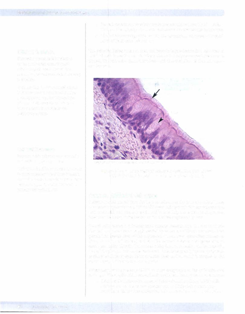
The epithelial lining of the trachea and bronchi is pseudostratified columnar in which all cells lie on the same basal membrane but only some reach the luminal surface. The only other place in the body with this epithelium is the male repro ductive tract.
Copyright McGraw-Hill Companies. Used with permission.
Figure 1-7-3. Pseudostratified columnar epithelium with goblet cells (arrowhead) surrounded by ciliated cells (arrow)
Tracheal Epithelial CellTypes
Columnar cells extend from the basal membrane to the luminal surface. These cells contain approximately 200 to 300 apical cilia per cell that are intermingled with microvilli. The cilia are motile and beat to help move the secreted mucus layer over the lining of the trachea and out of the respiratory system.
Goblet cells secrete a polysaccharide mucous material into the lumen of tra chea. Mucus production is supplemented by secretions of the submucosal mixed glands. The mucus layer of the respiratory system traps particulate substances (dust, bacteria, and viruses) and absorbs noxious water-soluble gases such as ozone and sulfur dioxide. The mucus sticky layer is moved by the beating cilia toward the pharynx where it is swallowed. This movement is known as the mu cociliary escalator system. Most material (dust and bacteria) is trapped in the mucus layer, and is removed and digested.
Pulmonary neuroendocrine (PNE) cells are comparable to the endocrine cells in the gut. These epithelial neuroendocrine cells have been given various names:
•APUD cells (Amino-Precursor-Uptake-Decarboxylase) , DNES cells (Diffuse Neuro Endocrine System) and K (Kulchitsky) cells. The granules in these neuroendocrine cells contain hormones and active
peptides, bombesin (gastrin-releasing factor), leu-enkephalin, sero tonin, and somatostatin. These cells occur in clusters and are often located at airway branch points.
•Brush cells may represent goblet cells that have secreted their products or intermediate stages in the formation of goblet or the tall ciliated cells. They have short microvilli on their apical surfaces. Some of these cells have synapses with intraepithelial nerves, suggesting that these cells may be sensory receptors.
Basal cells are stem cells for the ciliated and goblet cells. The stem cells lie on the basal membrane but do not extend to the lumen ofthe trachea. These cells, along with the epithelial neuroendocrine cells, are responsible for the pseudostratified appearance ofthe trachea.
BRONCHI
The bronchial tree forms a branching airway from the trachea to the bronchioles. When the primary bronchi enter the lung, they give rise to 5 secondary or lobar bronchi-3 for the right lung and 2 for the left. The 5 lobes are further subdivided into 10 tertiary or segmental bronchi in each lung, which form bronchopulmo nary segments.
Chapter 7 • Respiratory System
Clinical Correlate
The columnar and goblet cells are sensitive to irritation. The ciliated cells become taller, and there is an increase in the number of goblet cells and submucosal glands.
Intensive irritation from smoking leads to a squamous metaplasia where
the ciliated epithelium becomes a squamous epithelium. This process is reversible.
Clinical Correlate
Bronchial metastatic tumors arise from Kulchitsky cells.
CopyrightMcGraw-Hi/I Companies. Used with permission.
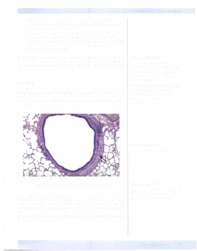
Figure 1-7-4. Bronchus with a plate of cartilage (arrow)
The epithelial lining of the bronchi is also pseudostratified. It consists of cili ated columnar cells, basal cells, mucus cells, brush cells and neuroendocrine
(APUD,gul DNES, or K) cells. There are also seromucous glands in the submucosa that empty onto the epithelial surface via ducts. The walls of bronchi contain ir re ar plates of cartilage and circular smooth-muscle fascicles bound together by elastic fibers. The number ofgoblet cells and submucosa glands decreases from the trachea to the small bronchi.
Clinical Correlate
Cystic fibrosis that results in abnormally thick mucus is in part due to defective chloride transport by Clara cells.
MEDICAL 81