Материал: Kaplan USMLE-1 (2013) - Anatomy
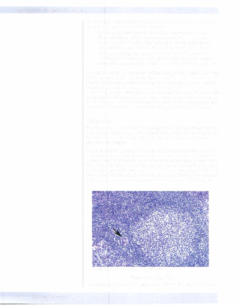

After passing through the white pulp, the central artery gives rise to smaller peni cillar arteries that enter the red pulp. Other radial artery and arteriolar branches of the central arteries empty into marginal located at the margin or outer zone of the white pulp, at its interface with red pulp. This provides an opportunity for blood-borne antigens to be removed from the blood by neighboring macro phages, making them available to antigen-processing cells, T and B lymphocytes of the white pulp, and initiating an immune response. These marginal sinuses eventually drain into splenic veins.
Chapter 6 • Immune Tissues
Note
95% of developingT cells die in the thymic cortex.
THYMUS
Like the lymph node, the thymus is divided into a distinct cortex and medulla. The thymus serves as a site ofmaturation ofT-lymphocyteprecursorsafter they exit from the bone marrow and arrive at the thymus via blood circulation. As these T cells develop, they are in intimate contact with epithelial cells of the thymus, also connected by desmosomes called dendritic epithelial cells because they have extensive thin branches oftheir cytoplasm extending out from the cell bodies. Because of the extensive branching of the epithelial cytoplasm there is extensive surface interaction possible between the developing T lymphocytes and thymic epithelial cells.
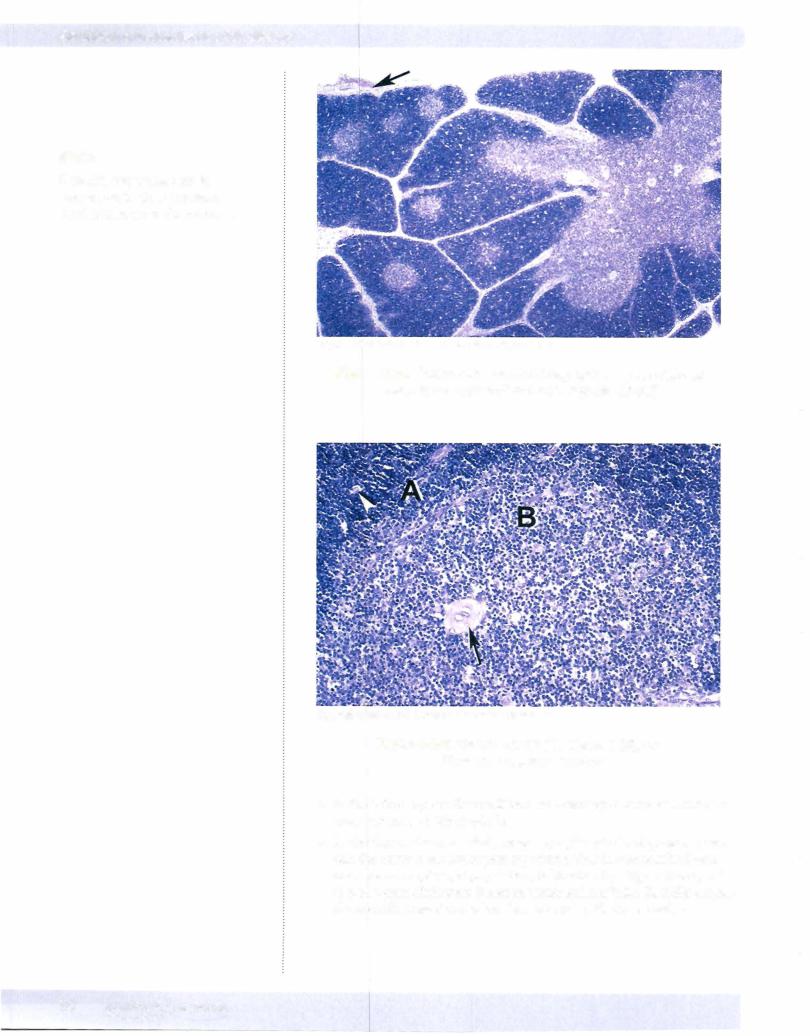
The thymic epithelial cells ofthe cortex are involved in clonal selection ofT cells. As the cells complete this process, the second set of dendritic epithelial cells in volved in clonal deletion of self-reactive T cells is located deeper in the medulla. The cortex is recognized by its darker staining. It has a high concentration of close-packed small lymphocytes with little cytoplasm and only a few epithelial cells and other cells. The medulla appears paler due to higher content of epithe lial and other cells with more cytoplasm separating the lymphocyte nuclei. Med ullary thymic epithelial cells form small aggregates of squamous epithelial cells called Hassall's corpuscles. Hassall's corpuscles are a reliable histologic marker for thymic medulla.
Another aspect ofthe thymus is that in the early stages ofT-cell maturation when positive and negative selection are taking place, there is a functional blood-thy mic barrier created by cortical thymic epithelial cells. These cells interact with the endothelium of thymic cortical blood vessels that "protects" the developing thymic lymphocytes from exposure to foreign antigens.
Note
The blood-thymus barrier consists of cortical thymic reticular cells joined by desmosomes, a dual basal lamina, and endothelial cells joined by tight junctions.
MEDICAL 73
Section I • Histology and Cell Biology
Note
Hassall's corpuscles secrete lymphopoietin that stimulates T-cell maturation in the medulla.
CopyrightMcGraw-Hill Companies. Used with permission.
Figure 1-6-8. Thymus with dark-staining cortex, lighter-staining medulla and connective tissue capsule (arrow)
Copyright McGraw-Hill Companies. Used withpermission.
Figure 1-6-9. Thymic cortex (A), medulla (B), and
Hassall's corpuscle (arrow)
•In the infant thymus there will be a well-developed outer cortex and a more central and pale medulla.
•In the thymus from an adult, most T-lymphocyte development is over and the thymus has undergone regression. This is characterized by a decrease in lymphocyte population, infiltration by adipose tissue, and loss of a clear distinction between cortex and medulla. Hassall's corpus cles are still present and often show central cystic degeneration.
74 MEDICAL

Chapter 6 • Immune Tissues
•In both infant and adult thymus there are prominent extensions of the fibrous capsule (trabecula) that extend deep into the thymus, separating incomplete lobules of thymic tissue. This is a distinctive feature of thymus compared to other lymphoid organs such as spleen and lymph node, as they are not as well divided into lobules.
•
•
•
ChapterSummary
The thymus contains trabeculae and has a cortical and medullary region.
Epithelial reticular cells and Hassall's corpuscles are located within the medulla. The cortex lacks germinal centers. The thymus protects developing T cells by the blood-thymus barrier.
The lymph node has 3 layers: outer cortial, inner cortical (paracortical), and medullary. The outer cortical layer contains most ofthe nodules and germinal centers. Most ofthe B lymphocytes reside here, whereas T lymphocytes reside in the paracortical layer. Dendritic cells within lymph nodes are antigen-presenting cells. High endothelial venules are the site of repopulation of lymph nodes and are located within the paracortical zones.
The spleen is very vascular and has red and white pulp. White pulp is composed of lymphoid tissue. T lymphocytes are located in the periarterial sheaths, while peripheral white pulp and germinal centers contain B lymphocytes. Red pulp consists of splenic cords and venous sinusoids. Its function is to delay passage of defective red blood cells to enable their elimination through phagocytosis by macrophages.
MEDICAL 75