Материал: Kaplan USMLE-1 (2013) - Anatomy
Section I • Histology and Cell Biology
Clinical Correlate
Chronic obstructive pulmonary disease (COPD) affects the bronchioles and includes emphysema and asthma.
•Emphysema is caused by a loss of elastic fibers and results in chronic airflow obstruction.
•Asthma is a chronic process characterized by a reversible narrowing of airways.
•Asthma is reversible; emphysema is not.
BRONCHIOLES
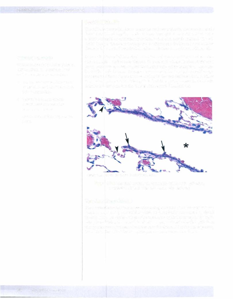
The wall ofabronchioledoes not contain cartilage or glands. The smooth-muscle fascicles are bound together by elastic fibers. The epithelium is still ciliated, but is a simple cuboidal or columnar epithelium rather than pseudostratified. The epi thelial lining of the airway is composed of ciliated cells (goblet and basal cells are absent in the terminal bronchioles) and an additional type called the Clara cell.
Clara cells (also called bronchiolar secretory cells) are nonciliated and secrete a serous solution similar to surfactant. They aid in the detoxificationofairborne toxins, and serve as a stem cell for the ciliated cells and for themselves. The num ber of Clara cells increases in response to increased levels of pollutants like ciga rette smoke. Clara cells are most abundant in the terminal bronchioles, where theymake up about 80 % ofthe epithelial cell lining; they are also involved with chloride ion transport into the lumens of the terminal bronchioles.
Copyright McGraw-Hill Companies. Used with permission.
Figure 1-7-5. Terminal bronchiole lumen (asterisk) with epithelium containing ciliated cells and Clara cells (arrows)
Terminal Bronchioles
The terminalbronchiole is the last conducting bronchiole. This bronchiole is fol lowed by respiratory bronchioles which are periodically interrupted by alveoli in their walls. The goblet cells are absent from the epithelial linings of the respi ratory bronchioles, but are still lined with a sparse ciliated cuboidal epithelium that prevent the movement of mucous into the alveoli. After the last respiratory bronchiole, the wall of the airway disappears and air enters the alveoli.
82 MEDICAL
Section I • Histology and Cell Biology
Clinical Correlate
Surfactant is synthesized after week 35 of gestation. Coricosteroids
induce the fetal synthesis of surfactant. High insulin levels in diabetic
mothers antagonize the effects of corticosteroids.
Infants of diabetic mothers have a higher incidence of respiratory distress syndrome.
84 MEDICAL
Copyright McGraw-Hill Companies. Used with permission.
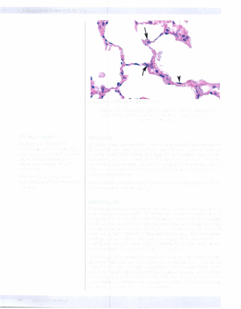
Figure 1-7-7. Alveoli with type I pneumocytes (arrowhead), type II pneumocytes (arrow), and alveolar macrophage (curved arrow) in the alveolar wall.
Surfactant
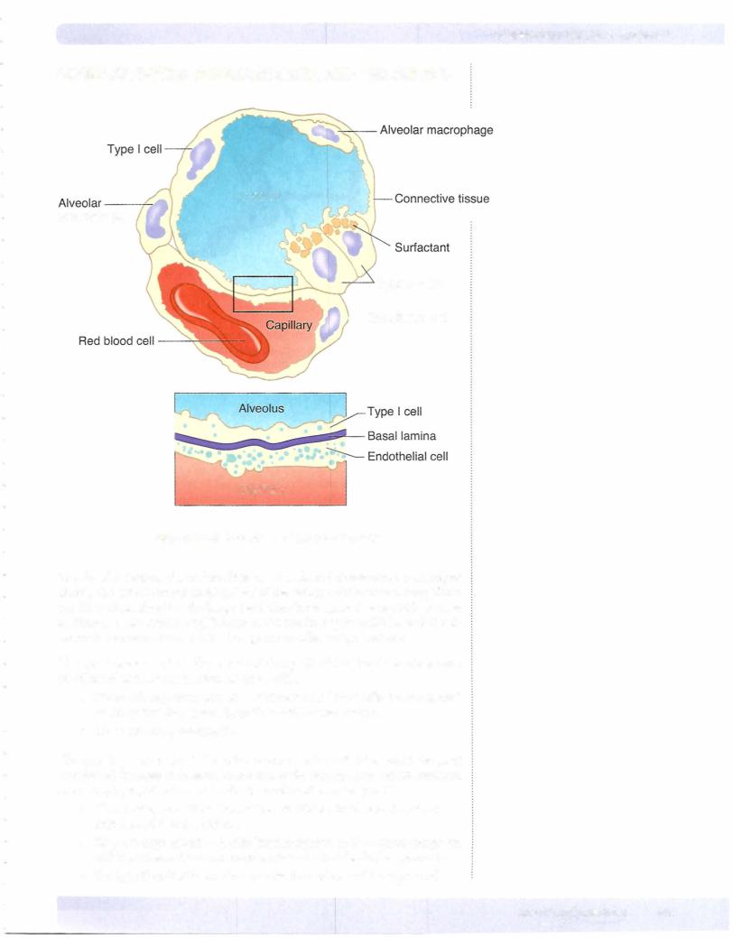
Surfactant is essential to maintain the normal respiratory mechanics ofthe alve oli. Production ofsurfactant in the fetus is essential for the survival ofthe neonate as it takes its first breath. Surfactant is composed of a mixture of phospholipids and surfactant proteins whose function is to aid in the spreading of the surfac tant at the alveolar air-water interface. The phospholipid in surfactant acts as a detergent which lowers the surface tension of the alveoli, and prevents alveolar collapse during expiration.
Most surfactant is recycled back to Type II cells for reutilization; some of it un dergoes phagocytosis by macrophages.
AlveolarWall
In the alveolarwallunder the alveolar epithelium is a rich network of capillaries arising from pulmonary arteries. The alveolar wall contains a variety of cells and extracellular fibers. The cells include fibroblasts, macrophages, myofibroblasts, smooth-muscle cells, and occasional mast cells. Type I and II collagens, as well as elastic fibers, are in the septa. Type I collagen is present primarily in the walls of the bronchi and bronchioles. Twenty percent of the mass of the lung consists of collagen and elastic fibers. Elastic fibers are responsible for the stretching and recoiling activities of the alveoli during respiration. These microscopic elements are responsible for the recoil of the lungs during expiration.
Gas exchange occurs between capillary blood and alveolar air across the blood gas barrier. This barrier consists ofsurfactant, the squamous Type I pneumocytes, a shared basal lamina, and capillary endothelium. The distance between the lu men of the capillary and the lumen of the alveolus can be as thin as 0. 1 microns. There are openings in the wall of most alveoli that from the poresofKohn. These pores are thought to be important in collateral ventilation. The diameter ofthese alveolar pores can be as large as l 0 to 15 microns.

Alveolar Macrophages
The alveolarmacrophages are derived from monocytes that exit the blood vessels in the lungs. The resident alveolar macrophages can undergo limited mitoses to form additional macrophages. These cells can reside in the interalveolar septa as well as in the alveoli. Alveolar macrophages that patrol the alveolar surfaces may pass through the pores of Kohn.
There are about 1 to 3 macrophages per alveolus. Alveolar macrophages vary in size from 15 to 40 microns in diameter. These macrophages represent the last defense mechanism of the lung. Macrophages can pass out of the alveoli to the bronchioles and enter the lymphatics or become trapped in the moving mucus layer and propelled toward the pharynx to be swallowed and digested. About 100 million lung macrophages are swallowed each day.
ChapterSummary
•The nasal cavities have 2 major areas: respiratory and olfactory.
•The respiratory area is lined by pseudostratified, ciliated columnar epithelium. Goblet cells are present as well. The olfactory area is in the posterosuperior area and contains bipolar neurons. Olfactory neurons are constantly replenished. Paranasal sinuses are located in the frontal, maxillary, ethmoid, and sphenoidal bones. They communicate with the nasal cavities. The nasopharynx is composed of stratified, squamous
nonkeratinized epithelium. The pharyngeal tonsil is an aggregate of nodular and diffuse lymphatic tissue within the posterior wall ofthe nasopharynx.
•Histologic features of the trachea, bronchi, and bronchioles are described in Table 1-7- 1 . Respiratory bronchioles contain alveoli and branch to form alveolar ducts, which terminate in alveolar sacs and are lined by squamous alveolar epithelium. Alveoli are terminal, thin-walled sacs of the respiratory tree responsible for gaseous exchange. They contain 2 kinds of cells.
-Type I cells provide a thin surface forgaseous exchange
-Type II cells produce surfactant.
Alveolar macrophages (dust cells) are located on the surface of alveoli and within the interalveolar connective tissue. They are derived from monocytes.
Chapter 7 • Respiratory System
Clinical Correlate
Alveolar macrophages have several other names: dust cells because they have phagocytosed dust or cigarette particles, and heart failure cells because they have phagocytosed blood cells that have escaped into the alveolar space during congestive heart failure.
MEDICAL 85