Материал: Kaplan USMLE-1 (2013) - Anatomy
Gastrointestinal System |
8 |
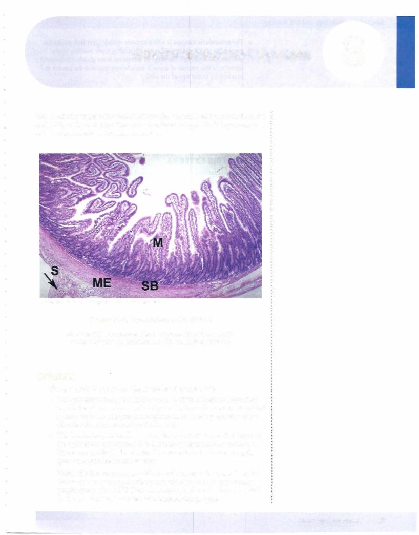
The alimentaryorgastrointestinal (GI) tract is a musculartubethatruns from the oral cavityto the anal canal. The GI tractwalls are composed of4 layers: mucosa, submucosa, muscularis externa, and serosa.
Copyright McGraw-Hill Companies. Used withpermission. GI
Figure 1-8-1. Organization of the tract
Mucosa (M) submucosa (SB), muscularis externa (ME), serosa or visceral peritoneum (S), mesentery (arrow)
MUCOSA
The mucosa is the innermost layer and has 3 components.
•The epithelium lining the lumen varies in different regions depending on whether the function is primarily conductive and protective (stratified squamous; in the pharynx and esophagus), or secretory and absorptive (simple columnar; stomach and intestine).
•The lamina propria is a layer of areolar connective tissue that supports the epithelium and attaches it to the underlying muscularis mucosae. Numerous capillaries form extensive networks in the lamina propria (particularly in the small intestine).
Within the lamina propria are blind-ended lymphatic vessels (lacteals) that carry out absorbed nutrients and white blood cells (particularly lymphocytes). The GALT (gutassociated lymphoid tissue), responsible for IgA production, is located within the lamina propria.
MEDICAL 87

Section I • Histology and Cell Biology
Clinical Correlate
Hirschsprung disease or aganglionic
megacolon is a genetic disease present in approximately 1 out of 5,000 live
births. It may result from mutations that affect the migration of neural crest cells into the gut. This results in a deficiency of terminal ganglion cells in Auerbach's plexus and affects of digestive tract motility, particularly in the rectum (peristalsis is not as effective and constipation results).
•The muscularis mucosa is a thin smooth-muscle layer that marks the inner edge of the mucosa. The muscle confers some motility to the mucosa and facilitates discharge of secretions from glands. In the small intestine, a few strands of smooth muscle may run into the lamina pro pria and up to the tips of the villi.
SUBMUCOSA
The submucosa is a layer of loose areolar connective tissue that attaches the mucosa to the muscularis externa and houses the larger blood vessels and mucus-secreting glands.
MUSCULARIS EXTERNA
The muscularis externa is usually comprised of 2 layers of muscle: an inner cir cular and an outer longitudinal. The muscularis externa controls the lumen size and is responsible for peristalsis. The muscle is striated in the upper third of the esophagus and smooth elsewhere.
SEROSA
The serosa is composed of a mesothelium (a thin epithelium lining the thoracic and abdominal cavities) and loose connective tissue and comprises the outermost membrane. In the abdominal cavity, the serosa surrounds each intestinal loop and then doubles to form the mesentery within which run blood and lymphatic vessels.
INNERVATION
The GI tract has both intrinsic and extrinsic innervation. The intrinsic inner vation is entirely located within the walls of the GI tract. The intrinsic system is capable of autonomous generation of peristalsis and glandular secretions. An interconnected network of ganglia and nerves located in the submucosa forms the Meissner's plexus and controls much of the intrinsic motility of the lining of the alimentary tract. Auerbach's plexus contains a second network of neuronal ganglia, and is located between the 2 muscle layers of the muscularis externa. All GI-tract smooth muscle is interconnected by gap junctions.
The extrinsic autonomic innervation to the GI tract is from the parasympathetic (stimulatory) and sympathetic (inhibitory) axons that modulate the activity of the intrinsic innervation. Sensory fibers accompany the parasympathetic nerves and mediate visceral reflexes and sensations, such as hunger and rectal fullness. Visceral pain fibers course back to the CNS with the sympathetic innervation. Pain results from excessive contraction and or distention of the smooth muscle. Visceral pain is referred to the body wall dennatomes that match the sympathetic innervation to that GI tract structure.
IMMUNE FUNCTIONS
The lumen of the GI tract is normally colonized by abundant bacterial flora. The majority of the bacteria in the body-comprisingB12 about 500 different species are in our gut, where they enjoy a rich growth medium within a long, warm tube. Most of these bacteria are beneficial (vitamins and K production, additional
88 MEDICAL
Chapter 8 • Gastrointestinal System
digestion, protection against pathogenic bacteria) but a few species ofpathogenic microbes appear at times. Our gut has defense mechanisms to fight these patho gens (GALT and Paneth cells).
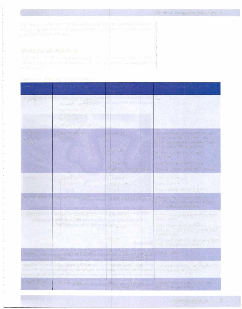
REGIONAL DIFFERENCES
Major differences lie in the general organization of the mucosa (glands, folds, villi,etc.) and in the types ofcells comprising the epithelia and associated glands
in the GI tract.
Table 1-8-1. Histology ofSpecific Regions
Region |
Major Characteristics |
Mucosal Cell Types at |
|
|
• |
|
Surface |
Esophagus |
Nonkeratinized stratified |
|
|
|
• |
squamous epithelium |
|
|
Skeletal muscle in |
|
|
|
|
muscularis externa |
|
|
|
(upper 1 /3) |
|
|
• Smooth muscle (lower 1/3) |
|
|
Stomach |
Rugae: shallow pits; |
Mucus cells |
|
(body and |
deep glands |
|
|
fundus) |
|
|
Chief cells |
Function ofSurface Mucosa! Cells
Secrete mucus; form protective layer against acid; tight junctions between these cells probably contribute to the acid barrier ofthe epithelium.
Secrete pepsinogen and lipase precursor
|
|
Parietal cells |
Secrete HCl and intrinsic factor |
|
|
Enteroendocrine (EE) cells |
Secrete a variety of peptide hormones |
Pylorus |
Deep pits; shallow, |
Mucous cells |
Same as above |
|
branched glands |
Parietal cells |
Same as above |
|
|
||
|
|
EE cells |
High concentration of gastrin |
Small intestine |
Villi, plicae, and crypts |
Columnar absorptive cells |
Contain numerous microvilli that |
|
|
|
greatly increase the luminal surface |
|
|
|
area, facilitating absorption |
Duodenum |
Brunner glands, which |
Goblet cells |
|
discharge alkaline |
|
|
secretion |
Paneth cells |
|
|
|
|
|
EE cells |
Secrete acid glycoproteins that protect mucosa! linings
Contains granules that contain lysozyme. May play a role in regulating intestinal flora
High concentration of cells that secrete cholecystokinin and secretin
Jejunum |
Villi, well developed plica, |
Same cell types as found in |
Same as above |
|
crypts |
the duodenal epithelium |
|
Ileum |
Aggregations of lymph |
M cells found over lym |
Endocytose and transport antigen from |
|
nodules called Peyer's |
phatic nodules and Peyer's |
the lumen to lymphoid cells |
|
patches |
patches |
|
Large intestine |
Lacks villi, crypts |
Mainly mucus-secreting |
Transports Na+ (actively) and water |
|
|
and absorptive cells |
(passively) out of lumen |
MEDICAL 89
Section I • Histology and Cell Biology
Oral Cavity
The epithelium of the oral cavity is a stratified squamous epithelium. Mucous and serous secretions of the salivary glands lubricate food, rinse the oral cav ity, moisten the foodfor swallowing and provide partial antibacterial protection. Secretions ofIgA from plasma cells within the connective tissue are transported through the gland epithelia to help protect against microbial attachment and in vasion.
Esophagus
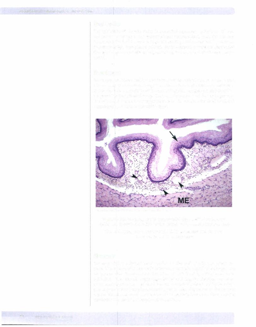
The esophagus is also lined by a stratified squamous epithelium. In thelowerpart ofthe esophagus there is an abrupt transition to the simple columnar epithelium of the stomach. Langerhans cells-macrophage-like antigen-presenting cells are present in the epithelial lining. The muscularis externa ofthe esophagus con sists ofstriated muscle in the upper third, smooth muscle in the distal third, and a combination ofboth in the middle third.
Copyright McGraw-Hill Companies. Used with permission.
Figure 1-8-2. Esophagus with non-keratinizing stratified squamous epithelium (arrow) and a thin lamina propriawith vessels (arrowheads)
The underlying muscularis externa (ME) is skeletal muscle from the upper half of the esophagus
Stomach
The stomach has 3 distinct histological areas: the cardia, body, and pyloric an trum. The mucosa ofthe stomach is thrown into folds (rugae) when empty, but disappears when the stomach is full. The surface is lined by a simple columnar epithelium. The stomach begins digestion by initiating the chemical and enzy matic breakdown ofingested food. Proteins are initially denatured by the acidic gastric juice before being hydrolyzed to polypeptide fragments by the enzyme pepsin. The chyme consists ofdenatured and partially broken-up food particles suspended in a semi-fluid, highly acidic medium.
90 MEDICAL