Материал: Kaplan USMLE-1 (2013) - Anatomy

Внимание! Если размещение файла нарушает Ваши авторские права, то обязательно сообщите нам

Chapter 10 • Male Reproductive System

ChapterSummary

•The testes contain seminiferous tubules and connective tissue stroma. Seminiferous tubules are the site of spermatogenesis. The epithelium contains Sertoli cells and spermatogenic cells.

-Sertoli cells synthesize androgen-binding protein and provide the blood­ testis barrier.

-Spermatogenic cells are germ cells located between Sertoli cells. They include spermatogonia, primary and secondary spermatocytes, spermatids, and spermatozoa.

Spermatozoa number about 60,000 per mm3 of seminal fluid. Each one has a head, which contains chromatin. Atthe apex ofthe nucleus is the acrosome. The tail contains microtubules.

•Interstitial cells of Leydig are located between the seminiferous tubules in the interstitial connective tissue. They synthesize testosterone and are activated by luteinizing hormone from the anterior pituitary.

•The genital ducts are composed oftubuli recti, rete testis, efferent ductules, ductus epididymis, ductus deferens, and ejaculatory ducts.

-Spermatozoa undergo maturation and increased motility within the ductus (vas) epididymis.

-Spermatozoa are stored in the efferent ductules, epididymis, and proximal ductus deferens.

•The urethra extends from the urinary bladderto the tip of the penis. The prostatic urethra is composed of transitional epithelium and the distal urethra of stratified epithelium.

•Seminal vesicles secrete alkaline, viscous fluid rich in fructose. They do not store spermatozoa.

•Secretions from the prostate gland are rich in citric acid, lipids, zinc, and acid phosphatase.

•Bulbourethral gland secretes mucous fluid into the urethra for lubrication priorto ejaculation.

•The penis is composed of 3 cylindrical bodies of erectile tissue: corpora cavernosa, corpus spongiosum, and trabeculae of erectile tissue. The corpora cavernosa is surrounded by the tunica albuginea.


 

137

MEDICAL

Chapter 11 • Female Reproductive System

Ovarian Follicles

An ovarian follicle consists ofan oocyte surrounded by one or more layers offol­ licular cells, the granulosa cells. In utero, each ovary initially contains 3 million primordial germ cells. Many undergo atresia as the number of follicles in a nor­ mal young adult woman is estimated to be 400,000. A typical woman willovulate only around 450 ova during her reproductive years. Allother follicles (with their oocytes) willfail to mature and willundergo atresia.

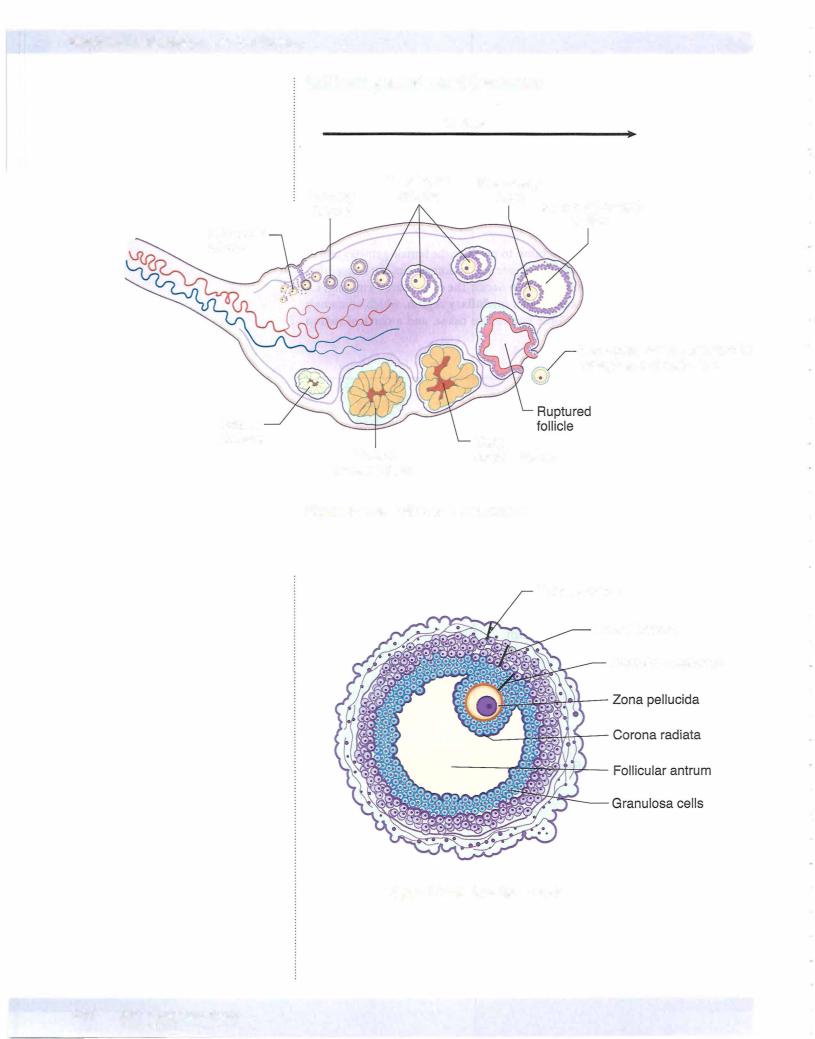
Before birth, primordial germ cells differentiate into oogonia that proliferate by mitotic division until they number in the millions. They all enter prophase ofthe first meiotic division in utero and become arrested (they are now designated as primordial follicles). The primordial follicles consist of a primary oocyte sur­ rounded by a single layer of squamous follicular cells, which are joined to one another by desmosomes.

Around the time of sexual maturity, the primordial follicles undergo further growth to become primary follicles in which the oocyte is surrounded by 2 or more layers of cuboidal cells. In each menstrual cycle after puberty, several primary follicles enter a phase of rapid growth. The oocyte enlarges and the surrounding follicular cells (now called granulosa cells) proliferate. Gap junc­ tions form between the granulosa cells. A thick layer of glycoprotein called the zona pellucida is secreted (probably by both the oocyte and granulosa cells) in the space between the oocyte and granulosa cells. Cellular processes of the granulosa cells and microvilli of the oocyte penetrate the zona pellucida and make contact with one another via gap junctions. Around this time the stroma surrounding the follicle differentiates into a cellular layer called the theca fol­ liculi. These cells are separated from the granulosa cells by a thick basement membrane. As development proceeds, 2 zones are apparent in the theca: the theca interna (richly vascularized) and the theca externa (mostly connective tissue). Cells of the theca interna synthesize androgenic steroids that diffuse into the follicle and are converted to estradiol by the granulosa cells.

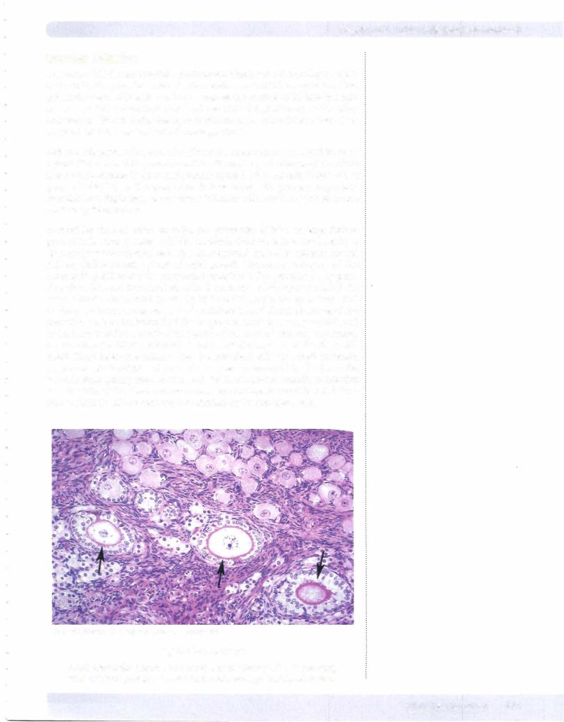
Copyright McGraw-Hill Companies. Used with permission.

Figure 1-1 1-4. Ovary

Small primordial follicles are at top and 3 primary follicles (arrows) with cuboidal granulosa cells and a thin zona pellucida are below.

MEDICAL 141

Источник: https://studfile.net/preview/14638320/