Материал: Kaplan USMLE-1 (2013) - Anatomy

Внимание! Если размещение файла нарушает Ваши авторские права, то обязательно сообщите нам

Chapter 11 • Female Reproductive System

Ovulation

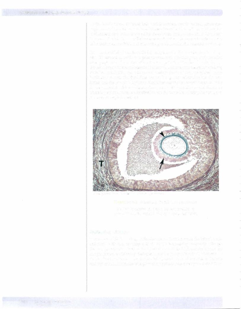
Ovulation occurs approximately mid-cycle andis stimulated by a surge ofluteiniz­ ing hormone secretedbythe anteriorpituitary. Ovulationconsists ofruptureofthe mature follicle andliberation ofthe secondaryoocyte (ovum)thatwillbe caughtby the infundibulum, the dilated distal end ofthe oviduct. The ovum remains viable for a maximum of24 hours. Fertilization most commonly occurs in the ampulla of the oviduct. Ifnot fertilized, the ovum undergoes autolysis in the oviduct.

Corpus Luteum

After ovulation, the wall ofthe follicle collapses and becomes extensively infold­ ed, forming a temporary endocrine gland called the corpus luteum. During this process the blood vessels and stromal cells invade the previously avascular layer of granulosa cells and the granulosa cells and those of the theca interna hyper­ trophy and form lutein cells (granulosa lutein cells and theca lutein cells). The granulosa lutein cells now secrete progesterone and estrogen and the thecalutein cells secrete androstenedione and progesterone. Progesterone prevents the devel­ opment ofnew follicles, thereby preventing ovulation.

In the absence ofpregnancy the corpus luteum lasts only 10-14 days. The lutein cells undergo apoptosis and are phagocytized by invading macrophages. The site ofthe corpus luteum is subsequently occupied by a scar of dense connective tis­ sue, the corpus albicans.

When pregnancy does occur, human chorionic gonadotropin produced by the placenta willstimulate the corpus luteum for about 6 months and then decline. It continues to secrete progesterone until the end ofpregnancy. The corpus luteum ofpregnancy is large, sometimes reaching 5 cm in diameter.

OVIDUCTS

The oviduct (Fallopian tube) is a muscular tube of about 12 cm in length. One end extends laterally into the wall ofthe uterus and the other end opens into the peritoneal cavitynext to the ovary. The oviduct receives the ovum from the ovary, provides an appropriate environment for its fertilization, and transports it to the uterus. The infundibulum opens into the peritoneal cavity to receive the ovum. Finger-like projections (fimbriae) extend from the end of the tube and envelop the ovulation site to direct the ovum to the tube.

Adjacent to the infundibulum is the ampulla, where fertilization usually takes place. A slender portion ofthe oviduct called the isthmus is next to the ampulla. The intramural segment penetrates the wall ofthe uterus.

MEDICAL 143

Section I • Histology and Cell Biology

Copyright McGraw-Hi/I Companies. Used with permission.

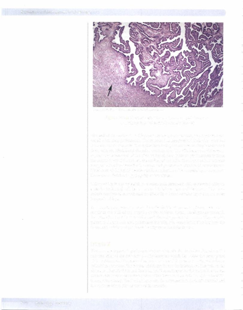
Figure 1-11-6. Oviduct with simple columnar epithelium and underlying layer of smooth muscle (arrow)

The wall of the oviduct has 3 layers: a mucosa, a muscularis, and a serosa com­ posed of visceral peritoneum. The mucosa has longitudinal folds that are most numerous in the ampulla. The epithelium lining the mucosa is simple columnar. Some cells are ciliated and the other are secretory. The cilia beat toward the uter­ us, causing movement of the viscous liquid film (derived predominantly from the secretory cells) that covers the surface of the cells. The secretion has nutrient and protective functions for the ovum and promotes activation of spermatozoa. Movement of the liquid together with contraction ofthe muscle layer transports the ovum or fertilized egg (zygote) to the uterus.

Ciliary action is not essential, so women with immotilecilia syndrome (Karta­ gener's syndrome) willhave a normal tubal transport of the ovum. The mus­ cularis consists of smooth-muscle fibers in a inner circular layer and an outer longitudinal layer.

An ectopic pregnancy occurs when the fertilized ovum implants, most com­ monly in the wall of the ampulla of the oviduct. Partial development proceeds for a time but the tube is too thin and the embryo cannot survive. The vascular placental tissues that have penetrated the thin wall cause brisk bleeding into the lumen of the tube and peritoneal cavity when the tube bursts.

UTERUS

The uterus is a pear-shaped organ that consists of a fundus which lies above the entrance sties of the oviducts; a body (corpus) which lies below the entry point of the oviducts and the internal os; a narrowing of the uterine cavity; and a lower cylindrical structure, the cervix, which lies below the internal os. The wall of the uterus is relatively thick and has 3 layers. Depending upon the part of the uterus, there is either an outer serosa (connective tissue and mesothelium) or adventitia (connective tissue). The 2 other layers are the myometrium (smooth muscle) and the endometrium (the mucosa ofthe uterus).

144 MEDICAL
Chapter 11 • Female Reproductive System

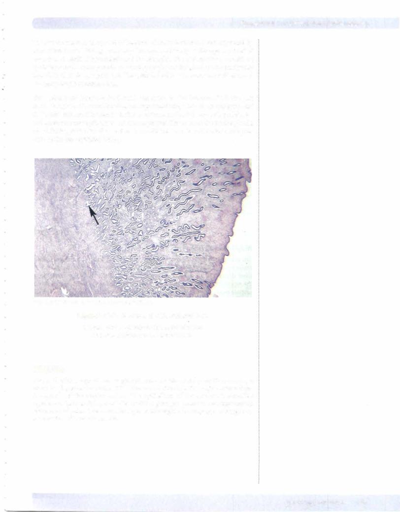
The myometrium is composed of bundles of smooth-muscle fibers separated by connective tissue. During pregnancy, the myometrium goes through a period of growth as a result of hyperplasia and hypertrophy. The endometrium consists of epithelium and lamina propria containing simple tubular glands that occasionally branch in their deeper portions. The epithelial cells are a mixture of ciliated and secretory simple columnar cells.

The endometrial layer can be divided into 2 zones. The functionalis is the part that is sloughed offat menstruation and replaced during each menstrual cycle, and the basalisis the portion retained after menstruation that subsequently proliferates and provides a new epithelium and lamina propria. The bases ofthe uterine glands, which lie deep in the basalis, are the source ofthe stem cells that divide and migrate to form the new epithelial lining.

CopyrightMcGraw-Hill Companies. Used with permission.

Figure 1-11 -7. Uterine wall with endometrium

Simple tubular glands to the right of arrow and myometrium to the left of arrow

VAGINA

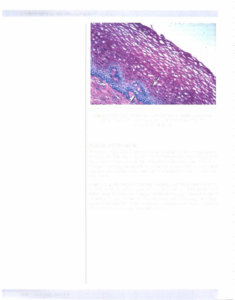
The wall of the vagina has no glands and consists of 3 layers: the mucosa, a muscular layer, and an adventitia. The mucus found in the vagina comes from the glands of the uterine cervix. The epithelium of the mucosa is stratified squamous. This thick layer of cells contains glycogen granules and may contain some keratohyalin. The muscular layer of the vagina is composed of longitudi­ nal bundles of smooth muscle.


MEDICAL

145

Section I • Histology and Cell Biology

CopyrightMcGraw-Hill Companies. Used with permission.

Figure 1-11-8. Vaginal epithelium with vacuolated stratified squamous epithelial cells that contain glycogen, which is removed during histological processing

MAMMARY GLANDS

The mammary glands are not part ofthe reproductive tract but are important ac­ cessory glands. The mammary gland is a tubuloalveolar gland consisting of 15-20 lobes drained by an equal number of lactiferous ducts that open at the tip of the nipple. The lobes are separated by connective tissue and varying amounts of adipose tissue. The histological structure varies according to sex, age, and physi­ ologic status.

At birth the gland consists only of short branching lactiferous ducts with no as­ sociated alveoli. As puberty approaches in the female, the ducts elongate and branch under the influence of ovarian hormones, and small spherical masses of epithelial cells appear at the ends ofthe branches. These cells are capable ofform­ ing functional acini in response to hormonal stimulation. Between the epithelial cells and the basal lamina are myoepithelial cells.

146 MEDICAL

Источник: https://studfile.net/preview/14638320/