Материал: Kaplan USMLE-1 (2013) - Anatomy

Внимание! Если размещение файла нарушает Ваши авторские права, то обязательно сообщите нам

Chapter 11 • Female Reproductive System

Copyright McGraw-Hill Companies. Usedwith permission.

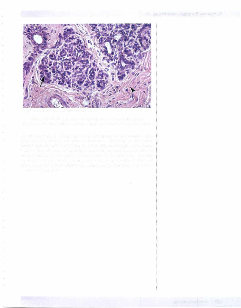
Figure 1-11-9. Breast tissue containing modified mammary gland tissue (arrow) surrounded by dense regular connective tissue (arrowhead)

The mammary glands enlarge significantly during pregnancy as a result of pro­ liferation ofalveoli at the ends ofthe terminal ducts. Alveoli are spherical collec­ tions of epithelial cells that become the active milk-secreting structures during lactation. The milk accumulates in the lumen ofthe alveoli and in the lactiferous ducts. Lymphocytes and plasma cells are located in the connective tissue sur­ rounding the alveoli. The plasma cell population increases significantly at the end ofpregnancy and is responsible for the secretion oflgA that confers passive im­ munity on the newborn.

MEDICAL 147

Section I • Histology and Cell Biology

Clinical Correlate

Breast cancer affects about 9% of women born in the United States. Most of the cancers (carcinomas) arise from epithelial cells ofthe lactiferous ducts.

ChapterSummary

•The female reproductive system is composed of ovaries, fallopian tubes, uterus, cervix, vagina, external genitalia, and mammary glands. The ovaries have 2 regions, the cortex and medulla. The former contain follicles and the latter vascular and neural elements. There are approximately 400,000 follicles at birth, of which approximately 450 reach maturity in the adult. The remaining follicles undergo atresia.

•Maturation involves the formation ofthe primary, secondary, and finally, the Graafian follicle. During ovulation, a rise in antral fluid causes the follicle to rupture. The ovum will degenerate in 24 hours unless fertilized by the spermatozoan. Following ovulation, the follicle changes in the following manner: theca interna cells become theca lutein cells and secrete estrogen;

while follicular cells become granulosa lutein cells, producing progesterone. If the ovum is fertilized, the corpus luteum persists for 3 months, producing progesterone. Its survival is dependent upon human chorionic gonadotropin secreted by the developing embryo. Thereafter, the placenta produces progesterone, required to maintain pregnancy.

•The fallopian tube is divided into the infundibulum, ampulla, isthmus, and interstitial segment. Fallopian tubes are lined by a mucosa containing cilia that beat toward the uterus, except in the infundibulum, where they beat toward the fimbria. Fertilization occurs in the ampulla, which is also the most frequent site of ectopic pregnancies.

•The uterus has 3 coats in itswall:

-The endometrium is a basal layer and superficial functional layer. The latter is shed during menstruation.

-The myometrium is composed of smooth muscle.

-The perimetrium consists of the peritoneal layer of the broad ligament.

The menstrual cycle results in cyclical endometrial changes. The first 3-5 days are characterized by menstrual flow. Thereafter, the proliferative stage commences. During this time, lasting 14 days, the endometrium regrows. This phase is estrogen-dependent. During the secretory phase, the endometrium continues to hypertrophy, and there is increased vascularity. This phase is progesterone-dependent. The premenstrual phase is marked

by constriction of spiral arteries leading to breakdown of the functional layer. Failure offertilization leads to a drop in progesterone and estrogen levels, and degeneration of the corpus luteum about 2 weeks after ovulation.

•The placenta permits exchange of nutrients and removal ofwaste products between maternal and fetal circulations. The fetal component consists of the chorionic plate and villi. The maternal component is decidua basalis.

Maternal blood is separated from fetal blood by the cytotrophoblast and syncytiotrophoblast.

•The vagina contains no glands. It is lined by stratified, squamous epithelium, rich in glycogen. During the estrogenic phase, its pH is acidic. During the postestrogenic phase, the pH is alkaline and vaginal infections could occur.
148 MEDICAL

Integument 12

GENERAL FEATURES

The integument consists of the skin (epidermis and dermis) and associated ap­ pendages (sweat glands, sebaceous glands, hairs, and nails). It is considered to be the largest organ in the body. The integument constitutes approximately 16% of total body weight. The integument functions to protect the body from injury, desiccation, and infection. It also participates in sensory reception, excretion, thermoregulation, and maintenance ofwater balance.

EPIDERMIS

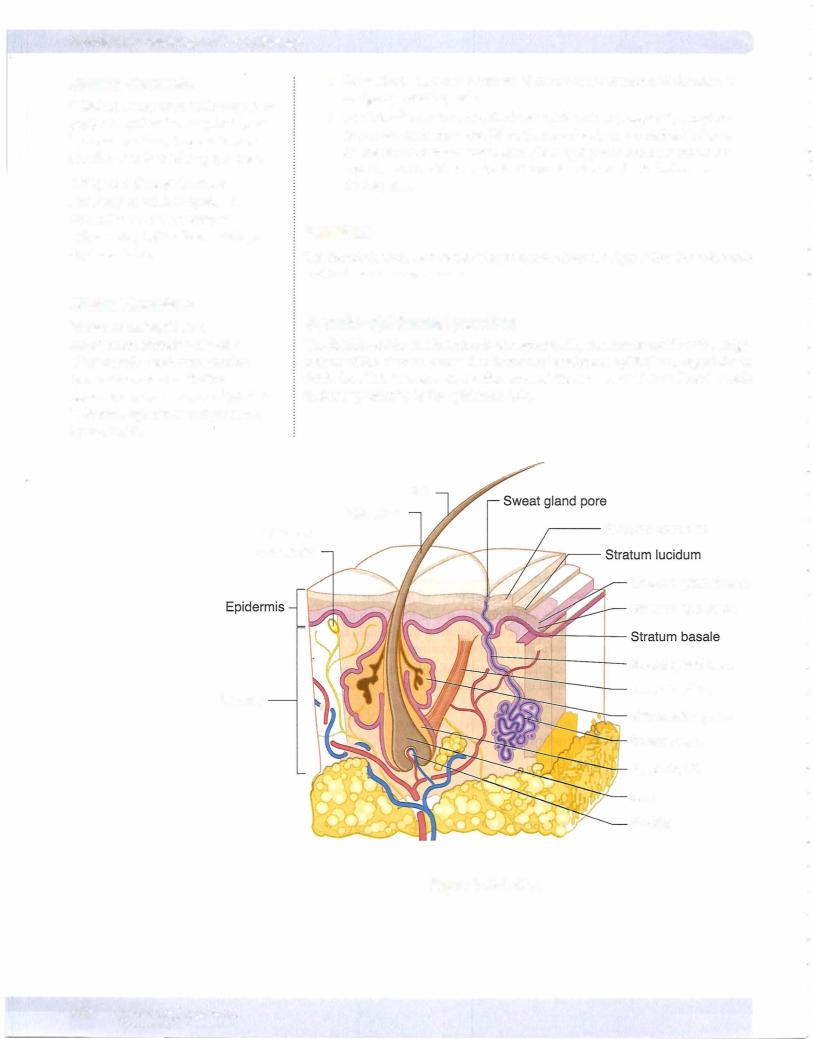
The epidermis is the outermost layer of the integument (Figure 1-12-1). It is a stratified squamous epithelial layer of ectodermal origin. It is devoid of blood vessels and consists of4 or 5 layers, from deep to superficial.

Layers

The layers of the epidermis are:

•Stratum basale (stratum germinativum) is a proliferative basal layer of columnar-like cells that contain the fibrous protein keratin.

•Stratum spinosum is a multilaminar layer of cuboidal-like cells that are bound together by means of numerous desmosomal junctions.

•Stratum granulosum consists of flat polygonal cells filledwith baso­ philic keratohyalin granules. Viewed at the electron microscopic level, these cells also contain numerous membrane-coating granules.

•Stratumlucidum is the transitional zone of flat eosinophilic or pale­ staining anucleated cells found only in regions with a thick stratum cor­ neum.

•Stratum corneum is the superficial stratum consisting of several layers of flat, anucleated, and cornified (keratinized) cells.

CellTypes

The epidermis contains several cell types:

•Keratinocytes are the most numerous and are responsible for the pro­ duction of the family ofkeratin proteins that provide the barrier func­ tion of the epidermis.

•Melanocytes are derivatives of neural crest ectoderm. They are found in the dermis and are also scattered among the keratinocytes in the basal layers of the epidermis. These cells produce the pigment melanin in the form of melanosomes that are transferred to keratinocytes.

Clinical Correlate

Pemphigus is an autoimmune blistering disorder caused by disruption of desmosomes linking keratinocytes.

Psoriasis results from an increase in the number of proliferating cells in stratum basale plus stratum spinosum. In addition, there is an increase in

the rate of cell turnover. This results in greater epidermal thickness and continuous turnover ofthe epidermis.

MEDICAL 149

Источник: https://studfile.net/preview/14638320/