Материал: Kaplan USMLE-1 (2013) - Anatomy

Внимание! Если размещение файла нарушает Ваши авторские права, то обязательно сообщите нам

Section II • Early Embryology

During the first 4 to 5 days ofthe first week, the zygote undergoes rapid mitotic division (cleavage) in the oviduct to form a blastula, consisting of increasingly smaller blastomeres. This becomes the morula (32-cell stage).

A blastocyst forms as fluid develops in the morula. The blastocyst consists ofan inner cell mass known as the embryoblast, and the outer cell mass known as the trophoblast, which becomes the placenta.

At the end of the first week, the trophoblast differentiates into the cytotropho­ blast and syncytiotrophoblast and then implantation begins (see below).

Clinical Correlate

Ectopic Pregnancy

Tubal

•The most common form of ectopic pregnancy; usually occurs when the blastocyst implants within the ampulla ofthe uterine tube because of delayed transport

•Risk factors: endometriosis, pelvic inflammatory disease (PID), tubular pelvic surgery, or exposure to diethylstilbestrol (DES)

•Clinical signs: abnormal or brisk uterine bleeding, sudden onset of abdominal pain that may be confused with appendicitis, missed menstrual period (e.g., LMP 60 days ago), positive human chorionic gonadotropin (hCG) test, culdocentesis showing intraperitoneal blood, positive sonogram

Abdominal

•Most commonly occurs in the rectouterine pouch (pouch of Douglas)

Implantation

The zona pellucida must degenerate for implantation to occur.

The blastocyst usually implants within the posterior wall ofthe uterus.

The embryonic pole ofblastocyst implants first.

The blastocyst implants within the functional layer of the endometrium during the progestational phase ofthe menstrual cycle.

Chapter Summary

•Fertilization occurs in the ampulla ofthe uterine tube with the fusion of the male and female pronuclei to form a zygote. During the first 4-5 days of

the first week, the zygote undergoes rapid mitotic division (cleavage) in the oviduct to form a morula before entering the cavity ofthe uterus.

•A blastocyst forms as fluid develops in the morula, resulting in a blastocyst that consists of an inner cell mass known as the embryoblast (becomes the embryo) and the outer cell mass known as the trophoblast (becomes the placenta). At the end ofthe first week, the trophoblast differentiates into the cytotrophoblast and syncytiotrophoblast and then implantation begins.

162 MEDICAL

Section II • Early Embryology

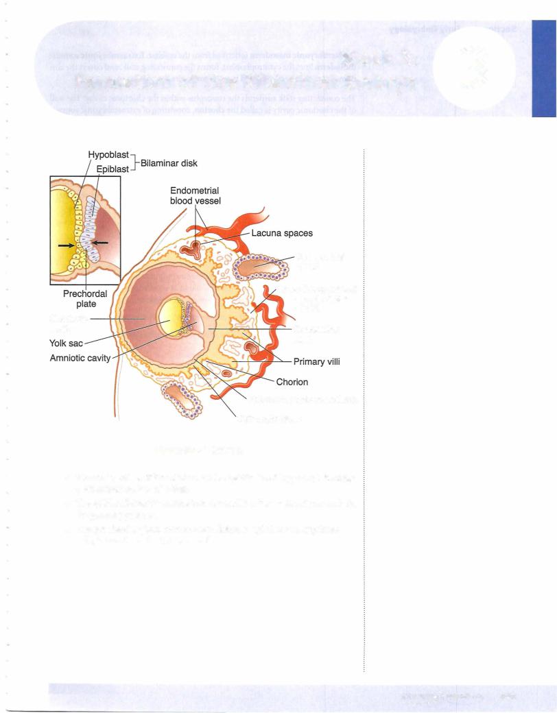
Extraembryonic mesoderm is derived from the epiblast. Extraembryonic somatic mesoderm lines the cytotrophoblast, forms the connecting stalk, and covers the am­ nion. Extraembryonic visceral mesoderm covers the yolk sac.

The connecting stalk suspends the conceptus within the chorionic cavity. The wall ofthe chorionic cavityis called the chorion, consisting ofextraembryonic somatic mesoderm, the cytotrophoblast, and the syncytiotrophoblast.

The syncytiotrophoblast continues its growth into the endometriurn to make con­ tact with endometrial blood vessels and glands. No mitosis occurs in the syncytio­ trophoblast. The cytotrophoblast is mitotically active.

Hematopoiesis occurs initially in the mesoderm surrounding the yolk sac (up to 6 weeks) and later in the fetal liver, spleen, thymus (6 weeks to third semester), and bone marrow.

Clinical Correlate

Human chorionic gonadotropin (hCG) is a glycoprotein produced by the syncytiotrophoblast. It stimulates progesterone production by the corpus luteum. hCG can be assayed in maternal blood or urine and is the basis for early pregnancy testing. hCG is detectable throughout pregnancy.

•Low hCG levels may predict a spontaneous abortion or ectopic pregnancy.

•High hCG levels may predict a multiple pregnancy, hydatidiform mole, or gestational trophoblastic disease.

ChapterSummary

•In the second week, implantation is completed with the rapid growth and erosion ofthe syncytiotrophoblast into the endometrium of the uterus where early utero-placental circulation is established. The inner cell mass of the first week differentiates into the epiblast and hypoblast cells and forms a bilaminar embryonic disk. An amniotic cavity develops from the epiblast and the primary yolk sac replaces the blastocyst cavity.

•The extraembryonic mesoderm and chorion are formed in the second week.

164 MEDICAL

Embryonic Period (Weeks 3-8)

4

Dorsal View

Cranial

Primitive node

8---

Caudal

A

Sectional View

Primitive node & streak

Epiblast (ectoderm)

-Amnion

BEndoderm

Figure 11-4-1 . Week 3

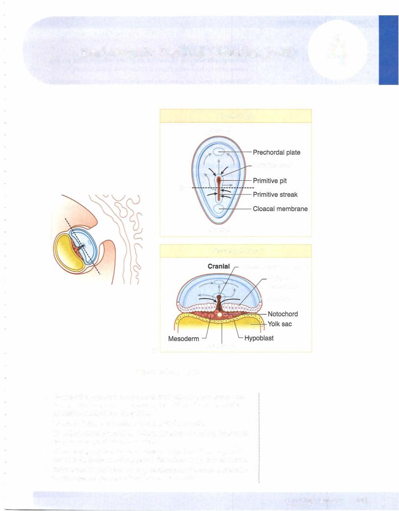
•Gastrulation--rocessp that produces the 3 primary germ layers: ecto­ derm, mesoderm, and endoderm; begins with the formation of the primitive streakwithin the epiblast

•Ectoderm forms neuroectoderm and neural crest cells.

•Mesoderm forms paraxialmesoderm (35 pairs of somites), intermedi­ ate mesoderm, and lateral mesoderm.

•Allmajor organ systems begin to develop during the embryonic period

(weeks 3-8).By the end of this period, the embryo begins to look human.

•Thirdweek: Gastrulation and early development of nervous and cardio­ vascular systems; corresponds to first missed period.

MEDICAL 165

Section II • Early Embryology

Clinical Correlate

Sacrococcygealteratoma: a tumor that arises from remnants ofthe primitive streak; often contains various types oftissue (bone, nerve, hair, etc)

Chordoma: a tumor that arises from remnants of the notochord, found either intracranially or in the sacral region

Hydatidiform mole: results from the partial or complete replacement ofthe trophoblast by dilated villi

•In a complete mole, there is no embryo; a haploid sperm fertilizes a blighted ovum and reduplicates so that the karyotype is 46,XX, with all chromosomes of paternal origin. In a partial mole, there is a haploid set of maternal chromosomes and usually 2 sets of paternal chromosomes so that the typical karyotype is 69,XXY.

•Molar pregnancies have high levels of hCG, and 20% develop into a malignant trophoblastic disease, including choriocarcinoma.

1 66 MEDICAL

Источник: https://studfile.net/preview/14638320/