Материал: Kaplan USMLE-1 (2013) - Anatomy

Внимание! Если размещение файла нарушает Ваши авторские права, то обязательно сообщите нам

Chapter 4 • Embryonic Period (Weeks 3-8)

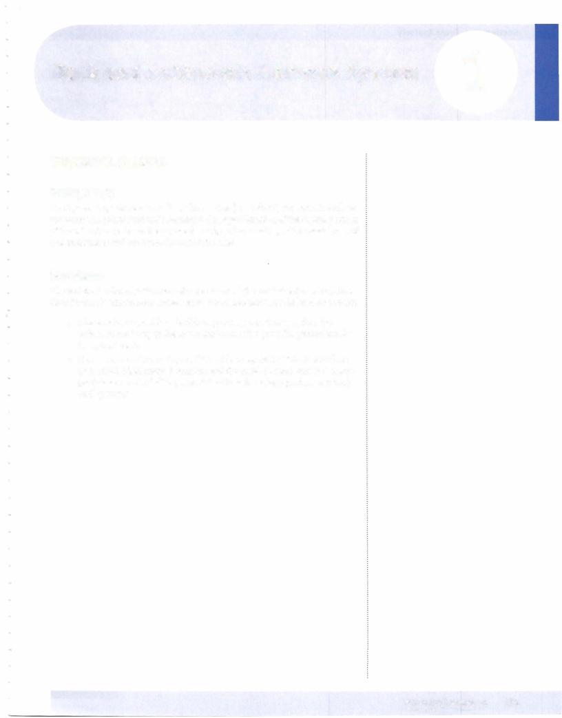
Table 11-4-1. Germ Layer Derivatives

Ectoderm

Mesoderm

Surface ectoderm

Muscle

Epidermis

Smooth

Hair

Cardiac

Nails

Skeletal


Inner ear, external ear

Connective tissue

Enamel ofteeth

All serous membranes

Lens ofeye

Bone and cartilage


Anterior pituitary (Rathke's pouch)

Blood, lymph, cardiovascular

Parotid gland

organs

Anal canal below pectinate line

Adrenal cortex


Neuroectoderm

Gonads and internal reproductive

organs

Neural tube


Central nervous system

Spleen


Retina and optic nerve

Kidney and ureter


Pineal gland

Dura mater

Neurohypophysis


Astrocytes


Oligodendrocytes


Neural crest ectoderm

Notochord

Nucleus pulposus

Adrenal medulla


Ganglia


Sensory-Pseudounipolar


Neurons


Autonomic-Postganglionic


Neurons


Pigment cells


Schwann cells


Meninges


Pia and arachnoid mater


Pharyngeal arch cartilage


Odontoblasts


Parafollicular (C) cells


Aorticopulmonary septum


Endocardial cushions


Yolk sac derivatives:


Primordial germ cells


Early blood cells and blood vessels

Endoderm

Forms epithelial lining of:

GI track: foregut, midgut, and hindgut

Lower respiratory system: larynx, trachea, bronchi, and lung

Genitourinary system: urinary bladder, urethra, and lower vagina

Pharyngeal pouches:

•Auditory tube and middle ear

•Palatine tonsils

•Parathyroid glands

•Thymus

Forms parenchyma of:

•Liver

•Pancreas

•Submandibular and sublingual glands

•Follicles ofthyroid gland

M EDICAL 167

Section II • Early Embryology

ChapterSummary

•The critical events ofthe third week are gastrulation and early development ofthe nervous and cardiovascular systems. Gastrulation is the process which establishes 3 primary germ layers that derive from epiblast: ectoderm, mesoderm, and endoderm. Gastrulation begins with the development of the primitive streak and node. The adult derivatives of ectoderm, mesoderm, and endoderm are given in Table 11-4-1.

168 MEDICAL

SECTION

GrossAnatomy

Back and Autonomic Nervous System

1

VERTEBRAL COLUMN

Embryology

During week 4, sclerotome cells ofthe somites (mesoderm) migrate medially to surround the spinal cord and notochord. After proliferation ofthe caudal portion ofthe sclerotomes, the vertebrae are formed, each consisting ofthe caudal part of one sclerotome and the cephalic part ofthe next.

Vertebrae

The vertebral column is the central component ofthe axial skeleton which func­ tions in muscle attachments, movements, and articulations ofthe head and trunk.

•The vertebrae provide a flexible support system that transfers the weight of the body to the lower limbs and also provides protection for the spinal cord.

•The vertebral column (Figure III- 1-1) is composed of 32-33 vertebrae (7 cervical, 12 thoracic, 5 lumbar, and the fused 5 sacral, and 3-4 coccy­ geal), intervertebral disks, synovial articulations (zygapophyseal joints) and ligaments.
MEDICAL 171

Источник: https://studfile.net/preview/14638320/