second sacral
Section Ill • Gross Anatomy
SPINAL MENINGES
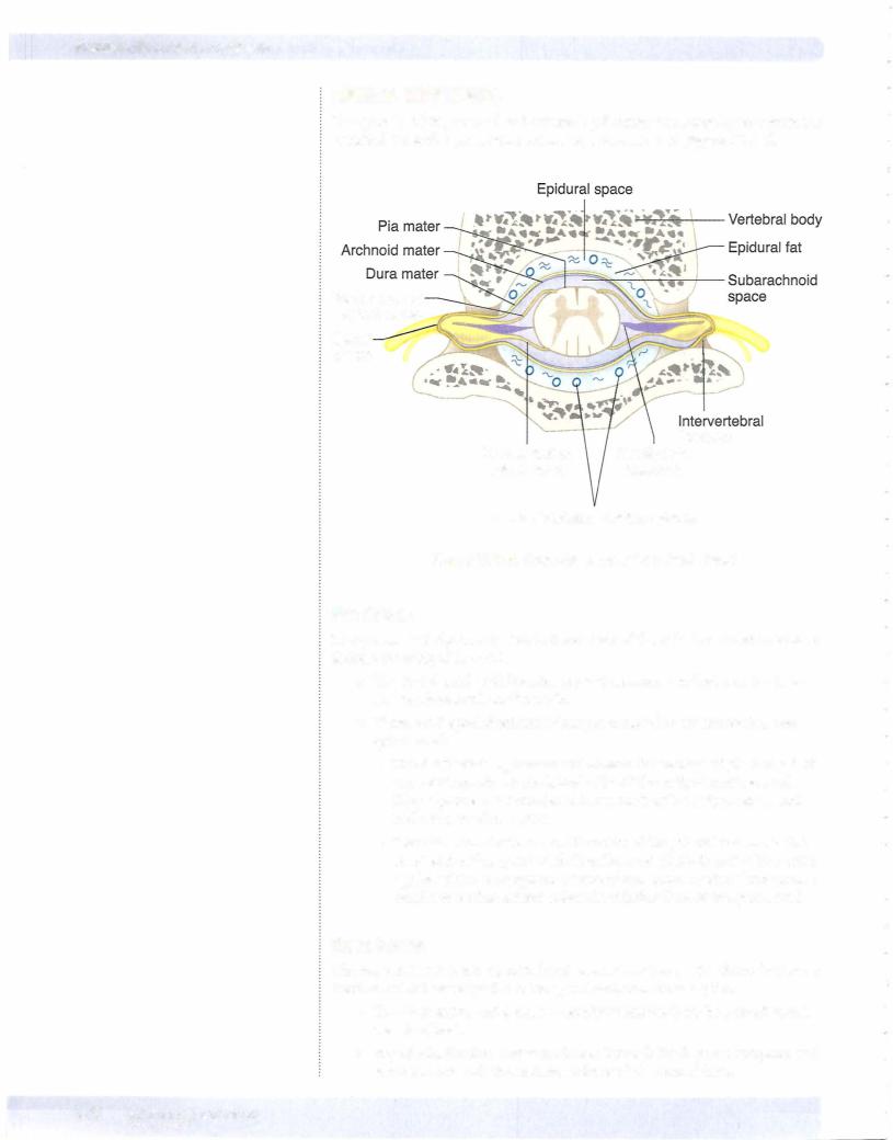
The spinal cord is protected and covered by 3 connective tissue layers within the vertebral canal: the pia mater, dura mater, and arachnoid (Figure III-1-5).
Ventral root of spinal nerve
Spinal nerve
Dorsal root of |
foramen |
Denticulate |
spinal nerve |
ligament |
Internal vertebral venous plexus
Figure 111-1-5. Cross-Section of Vertebral Canal
Pia Mater
The piamater is tightly attached to the surface of the spinal cord and provides a delicate covering of the cord.
•The spinal cord, with its covering of pia mater, terminates at the LI or
L2 vertebral levels in the adult.
•There are 2 specializations of the pia mater that are attached to the spinal cord:
-The denticulate ligaments are bilateral thickenings of pia mater that run continuously on the lateral sides of the midpoint of the cord. They separate the ventral and dorsal roots of the spinal nerves and anchor to the dura mater.
-The filum terminale is a continuation of the pia mater distal to the
lower end of the spinal cord. The filum terminale is part of the cauda equina which is composed of ventral and dorsal roots of lumbar and sacral nerves that extend below the inferior limit of the spinal cord.
Dura Mater
The dura mater is a tough, cylindrical covering of connective tissue forming a dural sac which envelops the entire spinal cord and cauda equina.
• The dura mater and dural sac terminate inferiorly at the vertebra level.
•Superiorly, the dura mater continues through the foramen magnum and is continuous with the meningeal layer of the cranial dura.