ChapterSummary
Vertebral Column
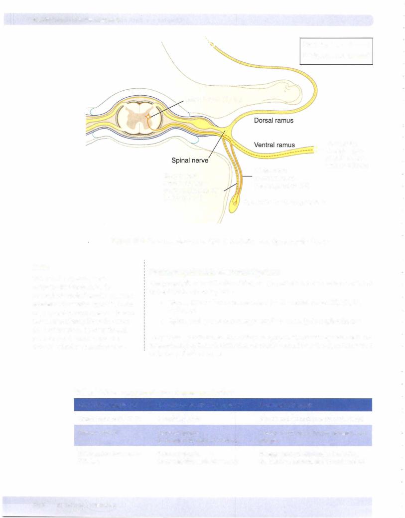
•The vertebral column is composed ofa series ofcervical, thoracic, lumbar, sacral, and coccygeal vertebrae connected by intervertebral disks and ligaments. The disks consist of an outer core of fibrocartilage, the annulus fibrosus, and an inner part-the nucleus pulposus-which develop from the notochord. Herniation ofthe nucleus pulposus is usually posterolateral where it can compress a spinal nerve at the intervertebral foramen.
•The spinal nerve exits the vertebral column at the intervertebral foramen. The foramen is bound superiorly and inferiorly by the pedicles of the vertebrae, anteriorly by the vertebral bodies and intervertebral disks, and posteriorly by the zygapophyseal joint.
•The spinal cord is covered by 3 protective layers of meninges: dura mater, arachnoid, and pia mater. The dura and dural sac terminate inferiorly at the second sacral vertebra, and the spinal cord terminates at the second lumbar vertebra. The cauda equina fills the lower part of the dural sac and contains the filum terminate and the ventral and dorsal roots ofthe lumbar
and sacral spinal nerves. Between the arachnoid and pia is the subarachnoid space that contains cerebrospinal fluid (CNS), and between the dura mater and the vertebrae is the epidural space, which contains fat and a plexus of veins. Spinal taps are performed at the level ofthe L4 vertebra (located at the horizontal level ofthe iliac crest) to avoid puncturing the spinal cord.
Autonomic NervousSystem
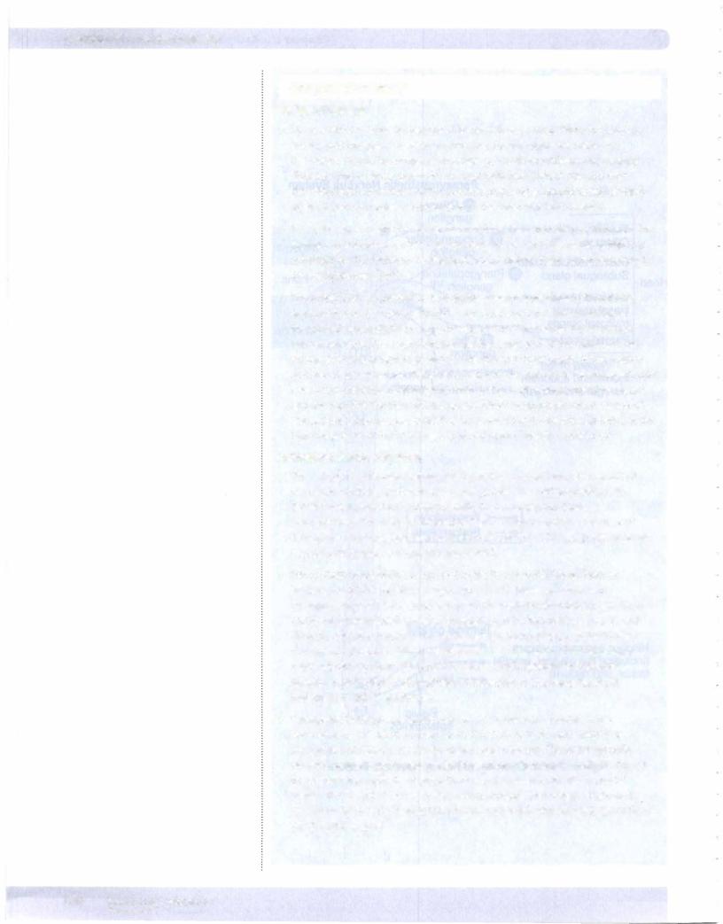
•The autonomic nervous system (ANS) provides visceral motor innervation to smooth muscle, cardiac muscle, and glands. The ANS is divided into
2 divisions: sympathetic (thoracolumbar) and parasympathetic (craniosacral). The peripheral distribution ofthese 2 divisions consists of
2 neurons: preganglionic neuron (cell bodies in the CNS) and postganglionic neuron (cell bodies in motor ganglia in PNS).
•Sympathetic preganglionic cell bodies are found in the lateral horn of the gray matter of spinal cord segments Tl-L2. These synapse with
postganglionic cell bodies located in either chain (paravertebral) ganglia or collateral (prevertebral) ganglia. Sympathetics to the body wall, head, and thoracic viscera synapse in the chain ganglia. Sympathetics to the foregut and midgut (thoracic splanchnic nerves: T5-T12) and to the hindgut and pelvic viscera (lumbar splanchnic nerves: L1-L2) synapse in collateral ganglia. Interruption of sympathetic innervation to the head results in ipsilateral Homer's syndrome.
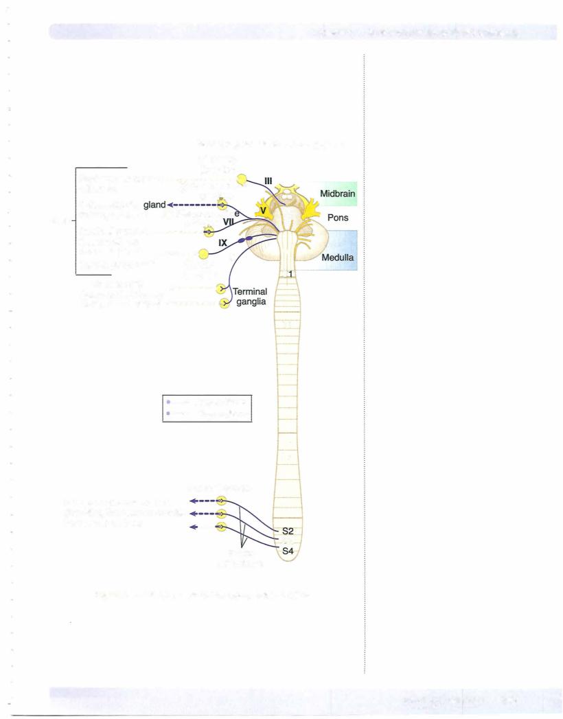
•Parasympathetic preganglionic neuron cell bodies are located in the brain stem nuclei of cranial nerves Ill, VII, IX, X, or in the gray matter of the spinal cord segments S2-S4 (pelvic splanchnics). The preganglionic
neurons synapse with postganglionic neurons in terminal ganglia scattered throughout the body. Parasympathetics to the head originate in cranial nerves Ill, VII, and IX; those to the thorax, foregut, and midgut originate in cranial nerve X; and those to the hindgut and pelvic viscera originate in the S2-S4 cord segments.