Section Ill • Gross Anatomy
AUTONOMIC NERVOUS SYSTEM
The autonomic nervous system (ANS) is concerned with the motor innervation of smooth muscle, cardiac muscle, and glands of the body.
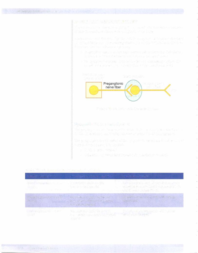
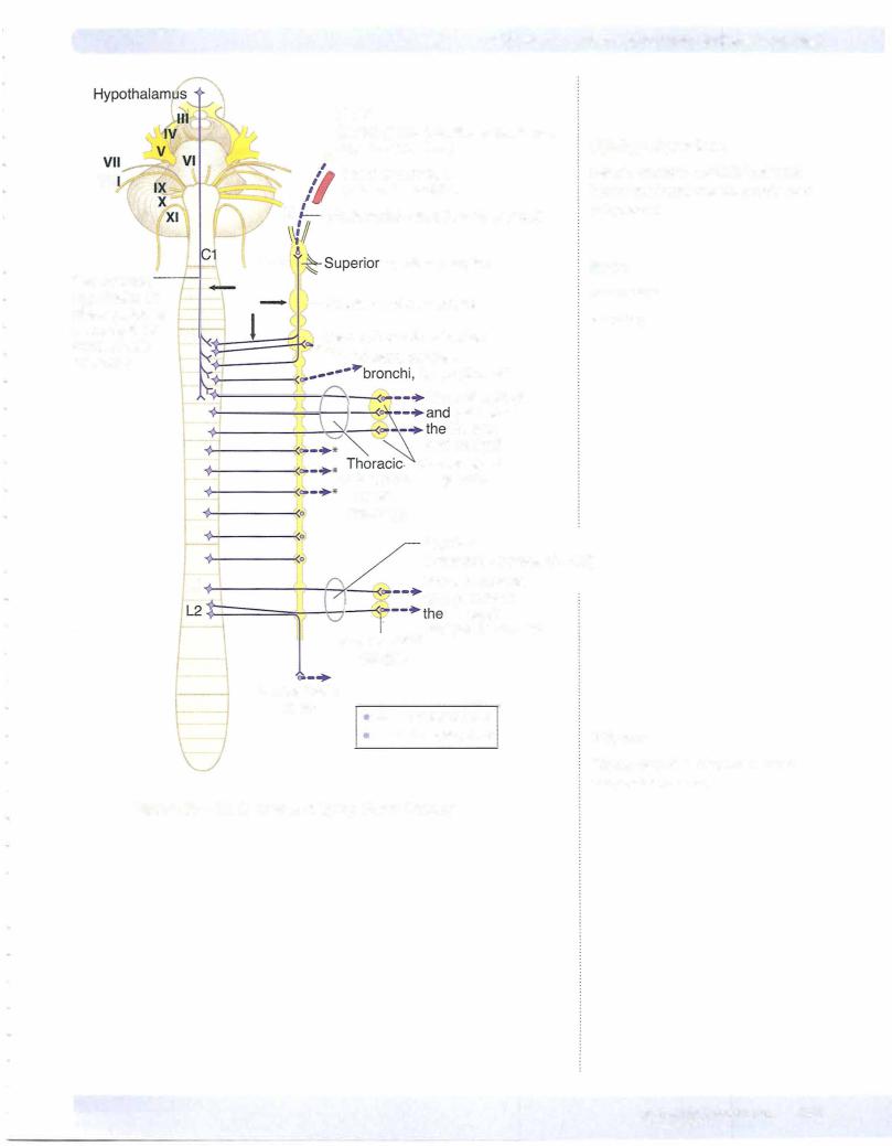
Anatomically and functionally, the ANS is composed of 2 motor divisions: ( 1 ) sympathetic and (2) parasympathetic (Figure III- 1 -9). In both divisions, 2 neurons formglian autonomic pathway.
• Pregan onicneuronshave their neuronal cell bodies in the CNS (formed by neuroectoderm); their axons exit in cranial and spinal nerves.
•Postganglionic neurons have cell bodies in autonomic ganglia in the peripheral nervous system (PNS) (formed by neural crest cells)
|
Central nervous |
Motor ganglion |
|
system (CNS) |
|
Postganglionic |
|
|
|
|
nerve fiber |
Target
Figure 111-1-9. Autonomic Nervous System
Sympathetic Nervous System
The preganglionic cell bodies of the sympathetic nervous system are found in the lateralhorn gray matter ofspinal cord segments TI-12 (14 segments).
The postganglionic cell bodies of the sympathetic system are found in one of
2types of motor ganglia in the PNS:
•Chain or paravertebral
•Collateral or prevertebral (found only in abdomen or pelvis)
Table 111-1-1. Sympathetic = Thoracolumbar Outflow |
|
Origin (Preganglionic) |
Site ofSynapse (Postganglionic) |
Innervation (Target) |
Spinal cord levels |
Sympathetic chain ganglia |
Smooth muscle, cardiac muscle and glands |
Tl-L2 |
(paravertebral ganglia) |
of body wall and limbs (Tl-L2), head (Tl-2) |
|
|
and thoracic viscera (Tl-5). |
Thoracic splanchnic nerves |
Prevertebral ganglia (collateral) |
Smooth muscle and glands ofthe foregut |
T5-T12 |
(e.g., celiac, aorticorenal, superior |
and midgut |
|
mesenteric ganglia) |
|
Lumbar splanchnic nerves |
Prevertebral ganglia (collateral) |
Smooth muscle and glands ofthe pelvic |
Ll-L2 |
(e.g., inferior mesenteric and pelvic |
viscera and hindgut |
|
ganglia) |
|