PLEURA AND PLEURAL CAVITY
Serous Membranes
Within the thoracic and abdominal cavities there are 3 serous mesodermal-de rived membranes which form a covering for the lungs (pleura), heart (pericar dium), and abdominal viscera (peritoneum).
Each ofthese double-layered membranes permits friction-reducing movements of the viscera against adjacent structures.
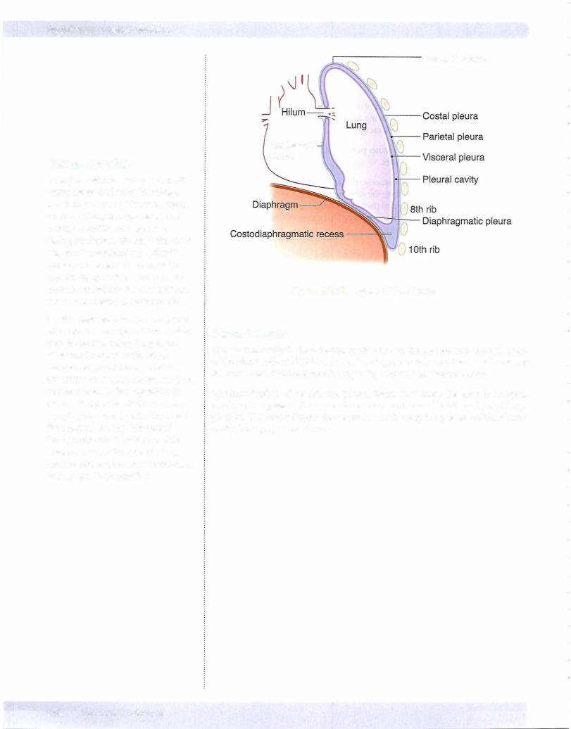
The outerlayer ofthe serous membranes is referred to as the parietallayer; and the inner layerwhich is applied directly to the surface ofthe organ, is calledthe visceral layer. The 2 layers are continuous and there is a potential space (pleural cavity) be tween the parietal and visceral layers containing a thin layer ofserous fluid.
Pleura
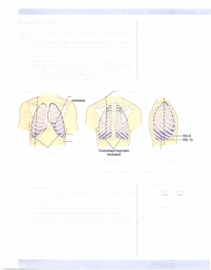
The pleura is the serous membrane that invests the lungs in the lateral compart ments of the thoracic cavity (Figure III-2-5). The external parietal pleura lines and attaches to the inner surfaces ofthe chest wall, diaphragm, and mediastinum. The innermost visceral layer reflects from the parietal layer at the hilum of the lungs and is firmly attached to and follows the contours ofthe lung. Visceral and parietal pleura are continuous at the root ofthe lung.
The parietal pleura is regionally named by its relationship to the thoracic wall and mediastinum (Figure III-2-5):
•Costal parietal pleura is lateral and lines the inner surfaces of the ribs and intercostal spaces
•Diaphragmatic parietalpleura lines the thoracic surface of the dia phragm
•Mediastinal parietalpleura is medial and lines the mediastinum. The mediastinal pleura reflects and becomes continuous with the visceral pleura at the hilum.
•Cervicalparietalpleura extends into the neck above the first rib where it covers the apex of the lung.
The visceral pleura tightly invests the surface of the lungs, following all of the fissures and lobes of the lung.
innervation of Pleura
The parietal pleura has extensive somatic sensory innervation provided by nerves closely related to different aspects of the pleura.
•The intercostal nerves supply the costal and peripheral portions of the diaphragmatic pleura.
•The phrenicnerve supplies the central portion of the diaphragmatic pleura and the mediastinal pleura.
The visceral pleura is supplied by visceral sensory nerves that course with the autonomic nerves.
Chapter 2 • Thorax
Clinical Correlate
Respiratory distress syndrome is caused by a deficiency of surfactant (type II pneumocytes). This condition is associated with premature infants, infants of diabetic mothers, and prolonged intrauterine asphyxia. Thyroxine and cortisol treatment increase the production of surfactant.
Surfactant deficiency may lead to hyaline membrane disease, whereby repeated gasping inhalations damage the alveolar lining. Hyaline membrane disease is characterized histologically by collapsed alveoli (atelectasis) and eosinophilic (pink) fluid covering the alveoli.
Clinical Correlate
Inflammation ofthe parietal pleural layers (pleurisy) produces sharp pain upon respiration. Costal inflammation produces local dermatome pain ofthe chestwallvia the intercostal nerves; whereby mediastinal irritation produces referred pain via the phrenic nerve to the shoulder dermatomes of C3-5.