Section Ill • Gross Anatomy
Clinical Correlate
A varicocele develops when blood collects in the pampiniform venous plexus and causes dilated and tortuous veins. This may result in swelling
and enlargement ofthe scrotum or enlargement ofthe spermatic cord above the scrotum. Varicoceles are more prominent when standing because of the blood pooling into the scrotum. A varicocele will reduce in size when the individual is horizontal.
Clinical Correlate
Cancers of the penis and scrotum will metastasize to the superficial inguinal lymph nodes, and testicular cancer will metastasize to the aortic (lumbar) nodes.
Clinical Correlate
In males, a cremasteric reflex can be demonstrated by lightly touching the skin of the upper medial thigh, resulting in a slight elevation of the testis. The sensory fibers of the reflex are carried by the Ll fibers of the ilioinguinal nerve and the motor response is a function ofthe genital
branch of the genitofemoral nerve that innervates the cremasteric muscle.
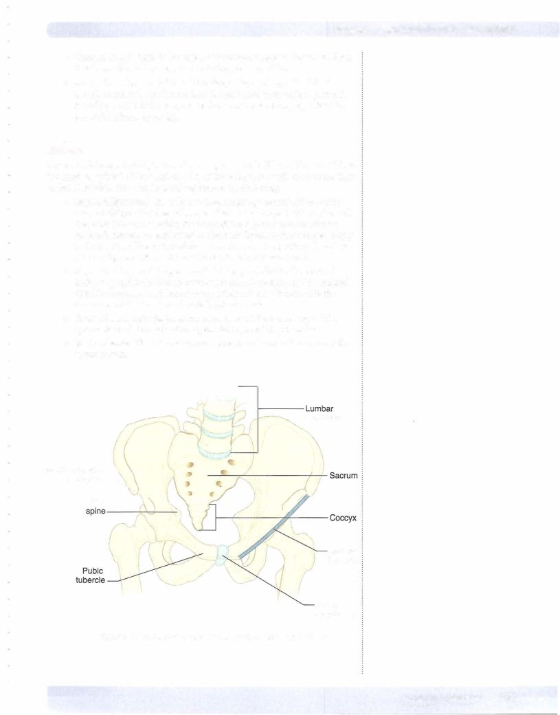
The entrance into the canal is the deep inguinal ring, located just lateral to the inferior epigastric vessels and immediately superior to the midpoint of the ingui nal ligament.
The superficial inguinal ring is the medial opening of the canal superior to the pubic tubercle.
Contents ofthe Inguinal Canal
1. Female Inguinal Canal
Round ligament ofthe uterus: The round ligament extends between the uterus and the labia majora and is a remnant of the gubernaculum that forms during descent of the ovary.
Ilioinguinal nerve (Ll ) is a branch of the lumbar plexus that exits the superficial ring to supply the skin of the anterior part of the mons pubis and labia majora.
2.Male Inguinal Canal
Ilioinguinalnerve (LI) is a branch of the lumbar plexus that exits the superficial ring to supply the skin of the lateral and anterior scrotum.
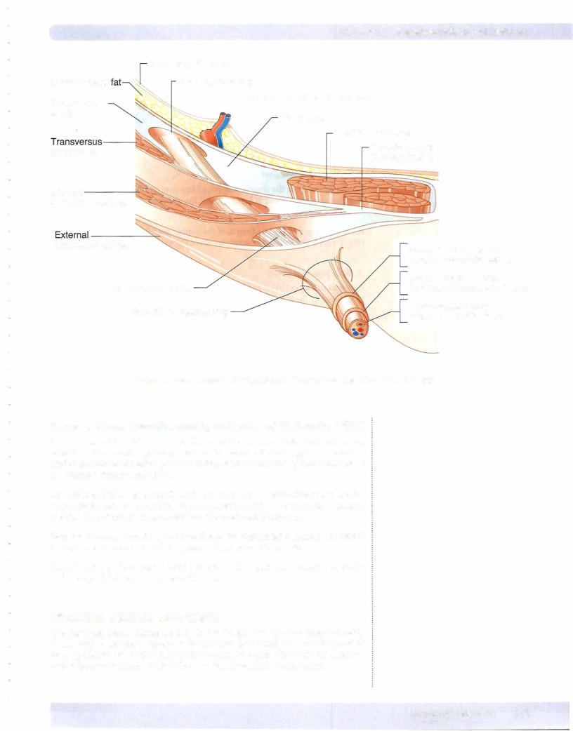
The spermatic cord is formed during descent of the testis and contains struc tures that are related to the testis. The cord begins at the deep ring and courses through the inguinal canal and exits the superficial ring to enter the scrotum. The spermatic cord is covered by 3 layers of spermatic fascia: external, middle, and internal. The cord contains the following:
•Testicular artery: A branch of the abdominal aorta that supplies the testis.
•Pampiniform venous plexus: An extensive network of veins draining the testis located within the scrotum and spermatic cord. The veins of the plexus coalesce to form the testicular vein at the deep ring. The venous plexus assists in the regulation of the temperature of the testis.
•Vas deferens (ductus deferens) and its artery
•Autonomic nerves
•Lymphatics: Lymphatic drainage of the testis will drain into the lumbar (aortic) nodes of the lumbar region and not to the superficial inguinal nodes which drain the rest of the male perineum.
Fascial Layers ofSpermatic Cord
There are 3 fascial components derived from the layers ofthe abdominal that sur round the spermatic cord (Figure III-3-2):
l.External spermatic fascia is formed by the aponeuroses of the external ab dominal oblique muscle at the superficial ring.
2.Middle or cremasteric muscle and fasciaare formed by fibers ofthe internal abdominal oblique within the inguinal canal The cremasteric muscle elevates the testis and helps regulate the thermal environment ofthe testis.
3.Internalspermaticfasciais formed by the transversalisfascia at the deep ring.