Boundaries of the Inguinal Canal
Roof
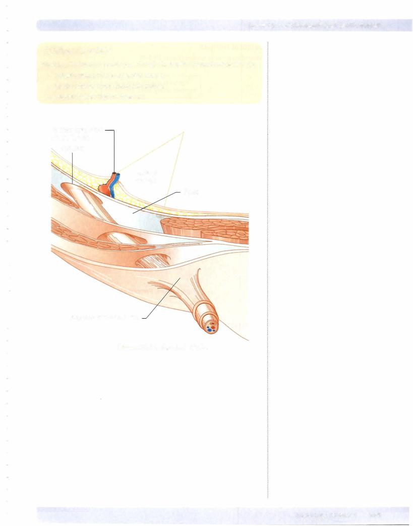
Formed by fibers ofthe internal abdominal oblique and the transverse abdominis muscles arching over the spermatic cord (Figure IIl-3-2)
AnteriorWall
Formed by aponeurosis ofthe external abdominal oblique throughout the ingui nal canal and the internal abdominal oblique muscle laterally
Floor
Formed by inguinal ligament throughout the entire inguinal canal and the la cunar ligament at the medial end
Posterior Wall:The posterior wall is divided into lateral and medial areas:
•Medialarea is formed and reinforced by the fused aponeurotic fibers of the internal abdominal oblique and transversus abdominis muscles
(conjoint tendon).
•Lateral area is formed bythe transversalis fascia and represents the weak area of the posterior wall.
•Inferior epigastric arteryandvein ascend the posterior wall just lateral to the weak area and just medial to the deep ring.
Descent ofthe Testes
The testis develops from the mesoderm of the urogenital ridge within the extra peritoneal connective tissue layer.
•During the last trimester, the testis descends the posterior abdominal wall inferiorly toward the deep inguinal ring guided by the fibrous gubernaculums.
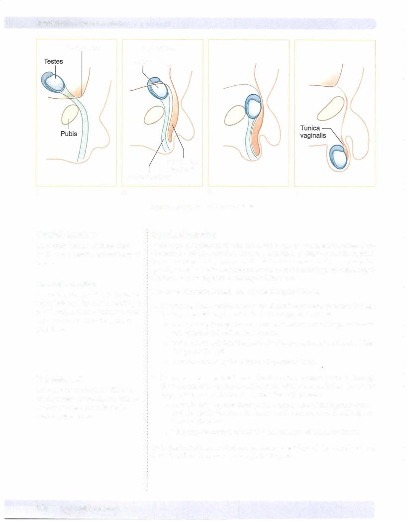
•An evagination of the parietal peritoneum and the peritoneal cavity extends into the inguinal canal called the processus vaginalis (Figure IIl-3-3). The open connection of the processus vaginalis with the perito neal cavity closes before birth.
•A portion of the processus vaginalis remains patent in the scrotum and surrounds the testis as the tunicavaginalis.